

Image source: Siemens Healthineers
News • Shape 23 event
Two new high-end MRI scanners for clinical and scientific use presented
Siemens Healthineers presented its two latest magnetic resonance tomographs designed for clinical and scientific use at the company's Shape 23 Keynote: Magnetom Cima.X with 3T field strength1 and Magnetom Terra.X with 7T.2
“Due to their high field strengths and strong gradient performance, both scanners will be optimal for detecting the finest structures in the body more clearly. By introducing AI-based algorithms on these high-end scanners for the first time, we reduce the scanning time in MRI by up to 50%3, while improving image quality," says Arthur Kaindl, Head of Magnetic Resonance Imaging at Siemens Healthineers.

Image source: Siemens Healthineers
The 3T Magnetom Cima.X will use the company's strongest gradient with an amplitude5 of 200 mT/m and a slew rate of 200 T/m/s. This is 2.5 times higher than the currently strongest MRI from Siemens Healthineers. Such a high gradient can help to better understand neurodegenerative diseases such as multiple sclerosis, even between relapses5. So-called microstructures can be visible more clearly with the help of the strong gradients5 and could play a major role in understanding these diseases. Microstructures can hardly be visualized with classical MR imaging. Thus, treatment could be initiated earlier, improving the outlook for patients.
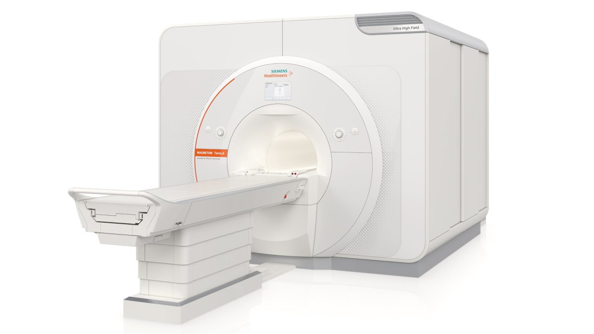
Image source: Siemens Healthineers
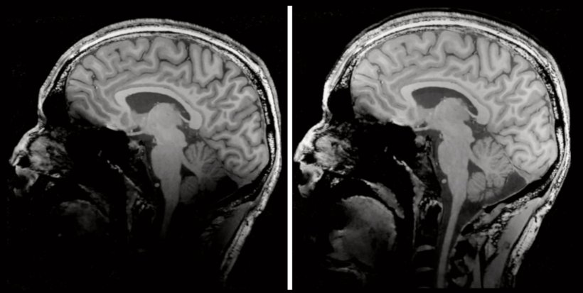
Magnetom Terra.X with a field strength of 7T represents the cutting edge of commercial MR imaging. The system will be the successor to Magnetom Terra, the first 7T clinical system, launched in 2017. The higher signal at 7T enables very high-resolution imaging for example of the head. This is particularly important in diseases where the detection of the smallest lesions can be crucial for the further treatment. Even in the field of knee imaging, the visibility of the smallest lesions can mean the decision for or against a surgery. The new hardware and software from Magnetom Terra.X, named "Ultra IQ technology" by Siemens Healthineers, significantly will increase the capabilities of a 7T system: MR imaging at these extremely high field strengths can lead to a darkening of the image impression for instance at the lower part of the head. With Magnetom Terra.X, potentially relevant findings are clearly visible even along the edges of the image. While classic MR imaging works especially well in body regions with a lot of water in the tissue, other substances in the body, such as sodium, can be better visualized with high field strengths. This form of imaging goes beyond the mere representation of anatomy and enables a visualization of processes in the body such as the metabolism.
Especially for research applications, both new scanners will offer extensive innovations, such as the new Open Recon platform6: "Open Recon allows us to run the reconstruction algorithms we have developed directly on the scanner. For the first time, we can transfer our research developments more easily, efficiently, and effectively into clinical routine," says Prof. Matthias Stuber from the University of Lausanne. In addition, both scanners will offer assistance systems that support scan preparation and the scan itself. Even these complex systems will be easy to operate for every user. The training effort will be minimal, and the scanners can be seamlessly integrated into the fleet.
References:
- Magnetom Cima.X is under development and is not commercially available. Future availability cannot be guaranteed.
- Magnetom Terra.X is under development and is not commercially available. Future availability cannot be guaranteed.
- Based on data acquired on Magnetom Cima.X. Data on file.
- >= 200 (+-3% for design tolerances)
- Huang, S. Y., Nummenmaa, A., Witzel, T., Duval, T., Cohen-Adad, J., Wald, L. L., & McNab, J. A. (2015). The impact of gradient strength on in vivo diffusion MRI estimates of axon diameter. NeuroImage, 106, 464–472.
- Open Recon is to add clinical reconstructions to the system, if signed and released for clinical use by SHS. Any other recon used e.g., by researchers is automatically labelled not for diagnostic use, which may require observation of national regulations
Source: Siemens Healthineers
16.11.2022


