News • Promising method for cancer imaging
Using photon counting for fast color X-ray images
New technology developed by researchers at the University of Houston could revolutionize medical imaging and lead to faster, more precise and more cost-effective alternatives to traditional diagnostic methods.

Image source: University of Houston
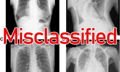
For years, doctors have relied on conventional 2D X-rays to diagnose common bone fractures, but small breaks or soft tissue damage like cancers often go undetected. More expensive and time-consuming MRI scans are not always suitable for these tasks in these detection or screening settings. Now, Mini Das, Moores professor at the University of Houston’s College of Natural Sciences and Mathematics and Cullen College of Engineering, has developed a 3D solution.
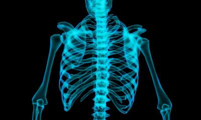
In a paper in the Journal of Medical Imaging, Das explains how photon counting detectors along with novel algorithms allow for more precise 3D visualization of different tissues and contrast agents by capturing X-rays at multiple energy levels simultaneously, which helps differentiate materials inside the body. “Right now, X-rays used in medical clinics and other industries collect incoming photons as a whole, similar to how white light contains all the colors, but they aren’t separated,” Das says. “So, while they can show differences in density — like distinguishing between bone and soft tissue — they can’t tell us exactly what materials are present.”
The photon counting detectors developed by Das’s team at UH can separate X-ray photons by their energy levels, similar to how a prism splits white light into different colors – and they can help identify specific materials, such as distinguishing between aluminum, plastic, iodine or other contrast agents like gadolinium used in medical imaging.
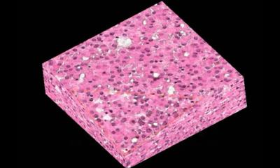
“This could improve cancer detection, for example,” Das says. “If you inject two different contrast agents – one targeting a tumor and another targeting inflammation – you could see where each one accumulates. Right now, we can see bright areas in an image, but we can’t always tell what they are. This technology would give us a much clearer, quantitative analysis. It would allow us to determine not just what’s inside an object, but what different materials are present and in what quantities.”
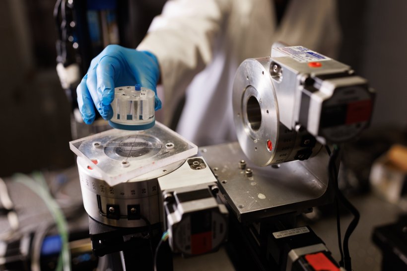
Image source: University of Houston
However, even with this advanced detection, some materials have similar X-ray properties, so distinguishing more than two or three at once can be a challenge. This is also amplified due to errors in the detectors as they separate photons by energy. But Das is working on a solution to that problem. “We have developed a method that compensates for these detector distortions by calibrating the detector using known materials,” Das says. “Once corrected, we can use the data along with the proposed novel algorithm, for accurate material decomposition – breaking down an image into its component materials. We do this in a multi-step solution from the same CT data collected improving accuracy.”
There are so many other potential applications for this technology including in materials imaging, baggage scanning for security, imaging for geophysics, and micro- and nano-electronics imaging — it’s very promising
Mini Das
Before the detectors can be widely used, there is still a lot of work to do. But Das says her team is working with industry partners in Europe to develop larger versions of these novel detectors and optimize their performance. “We’re still in the research and development phase,” Das says. “Right now, the detectors are small, and we need to refine their measurement accuracy. But once we solve those challenges, we can begin testing in real-world medical and industrial settings. There are so many other potential applications for this technology including in materials imaging, baggage scanning for security, imaging for geophysics, and micro- and nano-electronics imaging — it’s very promising.”
Previously, Das addressed a century-old problem in another innovative area related to the exploration of the wave nature of X-rays to significantly enhance soft material contrast. This research was featured in the scientific journal Optica last year.
Das’s research is funded through multiple agencies including NSF, CDMRP and NIH. The latest funding from the National Institute of Biomedical Imaging and Bioengineering aims to develop low-dose Micro-CT that utilizes multiple novel contrast mechanisms, thereby reducing radiation dose and imaging time which continues to be a significant issue.
Source: University of Houston
28.02.2025