Image source: LUMC
News • Single-cell metabolic profiling
Mass-spec and IMC give deep insights into tumors
Thanks to the use of highly advanced techniques, researchers at Leiden University Medical Center (LUMC) have succeeded in penetrating deeper into tumors than ever before.
They can now map the metabolism of individual cells in tumor tissue. This allows them to study not only which cells are present in a tumor, but also how they behave. Such knowledge is very valuable in the development of new therapies against cancer.
In the study, published in the scientific journal Nature Methods, the researchers describe a combination of two techniques that combined give a better idea of the so-called tumor environment: the set of cells, tissues and molecules that surround a tumor and interact with the tumor cells. Understanding this environment is crucial for developing more effective cancer therapies. But also to determine how dangerous a tumor is for a patient.
Over the years, metabolism has not received the attention it deserves. First, because it's very complex. And second because we didn't yet have the technology to get a grip on it, particularly in tissues
Noel de Miranda
Previously, researchers took tissues apart cell by cell. “The downside of that was that you no longer knew exactly where those cells came from. It was like taking apart a Lego structure and throwing it back into the box block by block,” said Marieke IJsselsteijn, one of the researchers. In the new study, the LUMC researchers used high-tech equipment that LUMC has in-house. They then combined imaging mass spectrometry (MS) and imaging mass cytometry (IMC) techniques. To do so, they worked intensively with LUMC's departments of pathology, human genetics, parasitology and the Center for Proteomics and Metabolomics.
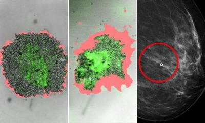
“With MS, we look at the metabolism of tumor tissue; how sugars are converted into energy, for example,” IJsselsteijn explains. “With IMC, we see how tumor cells, but also immune cells, behave in the tumor tissue. By combining these two techniques, we can see for each cell how active their metabolism is in the tissue and thus map much better what is going on in the tumor.”
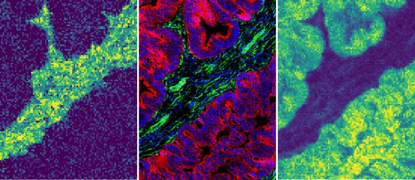
Image source: LUMC; adapted from: Nunes JB et al., Nature Methods 2024 (CC BY-NC-ND 4.0)
In cancer research, the metabolism of cancer cells plays an important role because it differs from the metabolism in healthy cells. Metabolism is essential to the body because it contains all the chemical processes that convert food into energy and building materials. Metabolism helps with growth, energy production, repair and breaking down waste products in our bodies.
IJsselsteijn: “From colon cancer, we know that in some of the tumors there are almost no immune cells. There doesn't seem to be that much going on. But based on research, we know that there is activity. By combining the two techniques, we hope to see, for example, whether there is certain activity in the tumor cells that prevents immune cells from getting there. Or that perhaps the immune cells have left because the nutrients have been absorbed by the tumor cells?”
Recommended article
Article • Clinical pathology
Focus on mass spectrometry
Mass spectrometry is pushing the boundaries of clinical pathology. Keep up-to-date with the latest research news, developments, and background information on the technique.
The researchers also hope to learn more by comparing tumors. “For example, you can have two tumors that look the same on the surface, while one tumor is much more aggressive than the other. We don't know why that is; we are missing something. Probably because we don't have all the information,” she says.
“Over the years, metabolism has not received the attention it deserves,” says Noel de Miranda, one of the researchers. “First, because it's very complex. And second because we didn't yet have the technology to get a grip on it, particularly in tissues. The development of this new technology allows us to understand the complexity of metabolism at the tissue level. This provides us with important information that we could not obtain before,” he says.
To visualize the metabolic system in a tissue with MS, fresh or frozen tumor tissue is used. The quality of such tissue deteriorates rapidly, making it previously difficult to match MS and IMC techniques. This has now been solved by fixing the tissue after determination via MS, that is, a thin layer of coating is added, keeping the tissue good for a very long time. The main outcome of this is that the scientists can now do research on the same piece of tissue twice in a row, without losing quality or damaging the tissue. “That gives us the ability to superimpose the two pictures given by the two techniques, allowing us to see not only which cells are all in a tissue, but also what they are doing. We are getting the complete picture. So in this case, 1 and 1 really is 3,” IJsselsteijn concludes.
Source: Leiden University Medical Center
27.09.2024