Image source: Postech
News • Contrast agent-free imaging of complex structures
Ultrafast ultrasound breaks new ground in imaging kidney vessels
High-speed video content can capture fleeting moments, such as bullets passing through glass. What if this technology were implement in ultrasound for medical examinations?
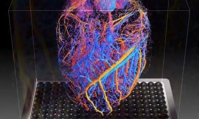
A research team at Postech (Pohang University of Science and Technology) has solved the mystery of kidney diseases using ultrafast ultrasound that captures 1,000 images in just one second. The research team has achieved imaging of the three-dimensional microvasculature of the kidneys using ultrafast ultrasound. Their technique is capable of visualizing the whole kidney microvasculature without any contrast agents. The findings were published as the inside back cover article of Advanced Science.

Image source: Postech
The kidney plays a role in filtering waste and eliminating unwanted substances from the bloodstream. Conditions such as hypertension and diabetes can compromise this vital function, leading to a kidney failure — irreversible condition necessitating lifelong treatment through artificial hemodialysis or donor kidney transplantation. Given the direct connection between blood perfusion in the kidneys and their filtration function, microvascular imaging can be a key indicator for both preventing and recovering from kidney failure.
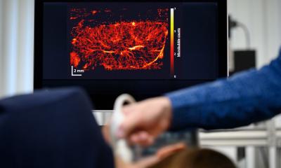
Representative contemporary medical imaging methods like CT (computed tomography) and MRI (magnetic resonance imaging) have limitations in capturing fine vascular structures due to their constraints in resolution and sensitivity. Moreover, the use of contrast agents in these methods is restricted due to the potentially fatal side effects in patients with kidney disease. In contrast, ultrasound imaging, considered safe enough for fetal monitoring, utilizes the Doppler effect to measure real-time blood flow velocity and direction without the need for contrast agents. However, the current imaging speed has limitations in capturing fine blood vessels with sufficient sensitivity. The research team has enhanced microvascular sensitivity by employing ultrafast ultrasound acquisition capturing 1,000 frames per second, a speed over 100 times faster than conventional ultrasound imaging.
Recommended article
Article • Diagnostic imaging
Ultrasound/Sonography
Ultrasound imaging is primarily known from prenatal care, but sonography is also used in many other places: it is just as indispensable for examining the thyroid and abdomen as it is in cardiology. Read all about application examples, new technology and more.
The system [...] has significant potential to be used to study blood circulation and functional impairment across various organs including the digestive system, circulatory system, and cerebral nervous system
Chulhong Kim
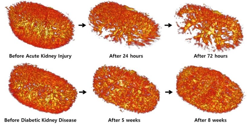
Using this technique, the researchers achieved a world-first by imaging the entire three-dimensional vascular network of renal artery, vein and 167μm (micrometer) thick interlobular arteries and veins in the renal cortex without the need for contrast agent. Furthermore, they conducted a continuous observation of renal vascular changes in an animal model induced with renal failure, performing multivariate analysis using hemodynamic and vascular morphological indicators. The results revealed a sharp decrease in renal blood flow during acute renal failure, while in the case of diabetic nephropathy, they identified chronic vascular degeneration in the kidneys accompanied by vascular distortion.
Professor Chulhong Kim explained, “The system allows us to understand the pathophysiology of diseases leading to kidney failure, enabling the observation of vascular changes before and after kidney transplantation.” He added, “It has significant potential to be used to study blood circulation and functional impairment across various organs including the digestive system, circulatory system, and cerebral nervous system.”
The research was supported by the National Research Foundation (NRF) grants, the Korea Medical Device Development Fund grant, the Korean Fund for Regenerative Medicine and BK21 FOUR projects (Pohang University of Science and Technology) funded by the Korean government (the Ministry of Science and ICT; the Ministry of Education; the Ministry of Trade, Industry and Energy; the Ministry of Health and Welfare).
Source: Pohang University of Science and Technology
20.01.2024