News • Neuro-monitoring
Thin-film electrodes reveal key insight into human brain activity
Thin-film electrodes developed at Lawrence Livermore National Laboratory (LLNL) have been used in human patients at the University of California, San Francisco, generating never-before-seen recordings of brain activity in the hippocampus, a region responsible for memory and other cognitive functions.
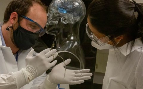
Photo: Julie Russell/LLNL
In a study published in the journal Nature Communications, surgeons at UCSF placed the flexible arrays on the brains of a group of patients while they were already undergoing epilepsy-related surgery. They recorded electrical signals across the exposed hippocampus while some patients were under anesthesia and others were awake, and conscious patients were given visual cues and spoke words while their neural activity was recorded. This approach allowed the researchers to detect traveling waves (TWs) moving across the hippocampal surface and identify new properties about them, including how they may contribute to human cognition. “We’ve developed an enabling technology for demonstrating a phenomenon that wasn’t really possible before,” said LLNL’s Implantable Microsystems Group Leader Razi Haque. “This challenge required creation of novel, conformable and higher-density electrodes that allows them to be more flexible and wrap around specific, deep regions of the brain. This study is validation that the approaches we’re using are getting us consistent, usable and useful data. That’s the driver for us as engineers — to be able to build the tools that scientists can use to do new science.”
LLNL developed the 32-channel multi-electrode arrays under the Defense Advanced Research Projects Agency (DARPA)-funded SUBNETS (Systems-Based Neurotechnology for Emerging Therapies) program, which aims to improve treatments for neuropsychiatric illnesses in military service members.
UCSF neurosurgeon and scientist Edward Chang, principal investigator of the SUBNETS program, speculated the arrays could work for a separate study examining the role of the hippocampus in memory function. By recording neural activity on the exposed hippocampal surface while patients were undergoing surgery, researchers could potentially confirm the existence of traveling waves, which scientists have long theorized play an important role in routing information used to form memories and perform other cognitive processing.
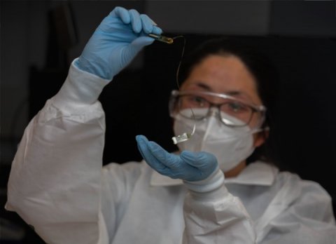
Photo: Julie Russell/LLNL
Previously, the nature of traveling waves in the human hippocampus has been controversial because previous in vivo studies have relied on penetrating depth electrode recordings. Those electrodes have provided researchers with only a few single-file recording sites in various layers of the hippocampus, making it nearly impossible to understand exactly how the waves are moving across the structure, according to the paper’s lead author and UCSF neurologist Jonathan Kleen.
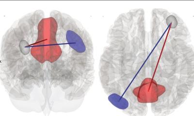
However, due to their high-density grid layout, small size (smaller than a dime) and their ability to conform to the hippocampal surface the LLNL-developed devices provided researchers with a critical “birds-eye-view” of how the signals moved and reversed over the surface like waves in water, Kleen said. “This new perspective helped us discover that traveling waves move both up and down the hippocampus,” Kleen said. “This ‘two-way street’ contrasts with the ‘one-way street’ previous neuroscience research has shown. This is a big deal because we believe this may be a fundamental mechanism of how the hippocampus acts as a major hub of information and memory processing for many other brain regions. In other words, the direction the wave is moving across the hippocampus may be a biomarker reflecting distinct neural processes as different circuits engage and disengage.”
The team used a machine learning approach to reveal that certain areas of the hippocampal surface activated more strongly depending on the direction the waves were moving. “This was further evidence that the route a wave is traveling may hint at what the hippocampus is up to at that moment,” Kleen said. Researchers noted that when one conscious patient tried to think of the name of a picture, traveling waves at one frequency consistently flowed toward the front of the structure. When the patient was awaiting the next trial, the waves reversed direction and flowed toward the back of the structure. The direction of wave travel may therefore reflect distinct cognitive processes when they occur, and potentially where information is flowing to support those processes, Kleen said.
The combination of precision data from these devices with next-generation data analytics promises to not only further our understanding of the inner workings of the brain but also lead to transformative cures for neurological disorders
Shankar Sundaram
The devices were built at LLNL and leverage knowledge gained over the course of more than a decade of research on thin-film micro-electrode arrays, beginning with the artificial retina project. LLNL engineers have improved the device’s processing steps through multiple fabrication test runs and design iterations, as well as years of bench-top tests to assess stability and performance, according to engineer Jenny Zhou, who fabricated the devices. “It definitely feels rewarding to know that our devices were tested in patients with success and enabled researchers to access new information to understand more about neural activity,” Zhou said of the recent study. “Kudos go to the interns, engineers, technicians who made it possible for us to continue the project. I started at LLNL as an intern working on the electrochemical side to characterize the electrode material that eventually became part of these thin-film devices, so for me personally, I’m glad to see it come full circle.”
Since the UCSF study concluded, LLNL engineers have doubled the number of electrodes on the flexible thin-film devices to 64 channels, enabling higher resolution sense and stimulation, and formed the arrays into a penetrating (or depth) probe. Engineers want to increase the channel count and density to hundreds, or even thousands, of electrodes per device. "The combination of precision data from these devices with next-generation data analytics promises to not only further our understanding of the inner workings of the brain but also lead to transformative cures for neurological disorders,” said Shankar Sundaram, director of LLNL’s Center for Bioengineering.
Source: Lawrence Livermore National Laboratory
15.05.2021


