Video • Imaging of speech disorder
MRI shows stuttering in real-time
Researchers at the University Medical Center Göttingen (UMG) and the Max Planck Institute for Multidisciplinary Sciences (MPI-NAT) have succeeded in visualizing the movement patterns of the internal speech muscles of a stuttering patient using real-time magnetic resonance imaging (MRI).
The method helps to improve our understanding of the mechanical aspects of stuttering, to identify muscle malfunctions in speech disorders, and to aid in the acquisition and reinforcement of new speech patterns. The results have been published in the Clinical Pictures section of The Lancet.
Stuttering is a speech disorder with abnormal movements and disturbed coordination of the movement dynamics of the internal speech muscles, the tongue and soft palate, which affects about 1% of the adult population. Until now this was poorly understood, since the muscles could not be observed from the outside, and it was not known just how they malfunctioned during stuttering. Recent advances in real-time MRI has now made it possible to visualize their movement patterns as they occur.

Image source: bvss/Michael Kofort
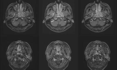
The collaboration of the "Interdisciplinary Working Group for Fluency Disorders" led by Prof. Dr. Martin Sommer in the Department of Neurology at the UMG, and the "Biomedical NMR" research group led by Prof. Dr. Jens Frahm at the MPI-NAT succeeded in making a video of the movements of the tongue and the soft palate of a 42-year-old stuttering patient while he was reading a text in the MRI scanner. For this video the MRI scanner had to provide 55 individual MRI scans per second. The video shows the movement patterns underlying the core symptoms of stuttering: involuntary repetitions of sounds and syllables, sound prolongations and audible or silent blocks. These show up in the video as sustained muscle contractions, i.e. a tightening of the muscles, and repetitive movements in parts of the tongue, lips and soft palate. Fluent speech segments were also detected, which are characteristic of this type of speech disorder.
These observations provide a better understanding of the incorrect functioning of the individual speech muscles and organs that occurs during stuttering. Translated to the clinic, the method will help to identify malfunctions in the movements of the speech muscles and organs in speech disorders, and aid in the acquisition and reinforcement of new speech patterns.
Prof. Sommer is convinced that: "By showing us the mechanical origin of the symptoms, real-time MRI improves our understanding of stuttering and will be an important tool in further research. And by enabling us to directly see where the internal speech muscles and organs make "wrong" movements, this method will also assist us greatly in the treatment of this multifaceted neuromuscular disorder."
Source: University Medical Center Göttingen
06.05.2024


