Image source: RSNA; from: Dirrichs et al., Radiology 2023
News • Congenital heart defect diagnosis
Photon-counting CT improves cardiac imaging in babies
A new advanced form of CT imaging called photon-counting computed tomography (PCCT) offers better cardiovascular imaging quality at a similar radiation dose compared to dual-source CT (DSCT) in infants with suspected cardiac heart defects, according to a new study.
The findings were published in Radiology, a journal of the Radiological Society of North America (RSNA).
Congenital heart defects are the leading cause of morbidity and mortality in the neonatal period, occurring in up to one percent of live births. Of those, approximately 25% are critical defects requiring surgical intervention within the first month after birth. A comprehensive assessment, including ultrasound, MRI and CT exams, is typically needed to plan for surgery and to create virtual and printed 3D reconstructions of the heart.

Image source: RSNA
“Infants and neonates with suspected congenital heart defects are a technically challenging group of patients for any imaging method, including CT,” said Timm Dirrichs, M.D., senior physician and specialist in cardiothoracic radiology in the Department of Diagnostic and Interventional Radiology at RWTH Aachen University Hospital in Aachen, Germany. “There is a substantial clinical need to improve cardiac CT of this vulnerable group. It’s essential to carefully map the individual cardiac anatomy and possible routes of surgical intervention using the highest possible diagnostic standards.”
PCCT is an emerging imaging technique that counts the exact number and measures the energy of incoming x-ray photons. Compared with DSCT technology, PCCT offers higher image resolution and/or reduced radiation doses, which is of particular interest when imaging children. The PCCT technique has already been shown to improve cardiovascular CT imaging in adults. However, data on neonates and small children are lacking.
“Our aim was to evaluate the image quality of first-generation photon-counting CT for cardiac imaging in children with suspected cardiac heart defects compared with third-generation dual-source CT (DSCT) and to compare the respective radiation exposure,” Dr. Dirrichs said.
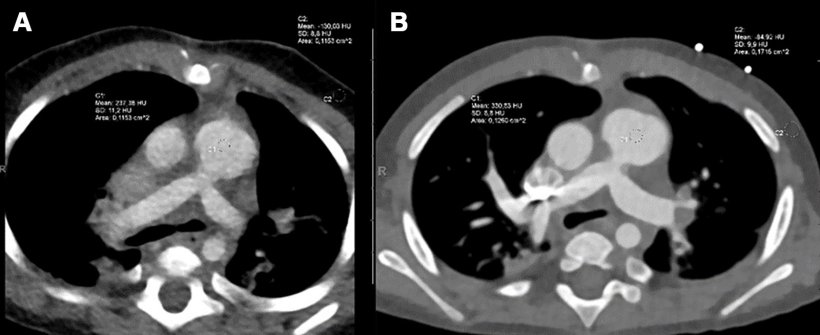
Image source: RSNA; from: Dirrichs et al., Radiology 2023
The research team analyzed existing clinical CT exams of 113 children who underwent contrast enhanced PCCT (30 infants), DSCT (83 infants) or both PCCT and DSCT (one infant) of the heart and thoracic aorta between January 2019 and October 2022. The study group consisted of 55 girls/58 boys (median age 66 days).
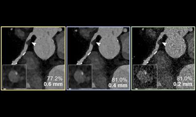
The researchers found that the PCCT images were sharper, with less image noise and greater contrast than DSCT images. The mean overall visual image quality ratings were higher for PCCT versus DSCT at a similar radiation dose. More than 97% of the PCCT images were at least diagnostic quality, compared to 77% of the DSCT images. “In our study, none of the PCCT examinations exhibited a poor image quality, and only a few were of limited or moderate quality,” Dr. Dirrichs said. He noted that of the DSCT images, almost one-quarter were of limited or non-diagnostic quality, and 40% were of moderate quality. “PCCT is a promising method that may improve diagnostic image quality and efficiency compared to DSCT imaging,” Dr. Dirrichs said. “This higher efficiency can be used to reduce the radiation dose at a given image quality level or to improve image quality at a given radiation level.”
Source: Radiological Society of North America
24.05.2023