Article • Diagnosis, prognosis, prediction
AI offers advances in cardiovascular imaging
Artificial Intelligence (AI) is providing numerous opportunities across clinical care in the field of cardiovascular imaging. While challenges remain, AI is being applied in terms of diagnosis and prognosis, defining cardiovascular imaging pathways, and image acquisition and analysis. It can also help cardiologists predict which patients may do well, or which treatments are best applied to those suffering from specific conditions.
Report: Mark Nicholls
Three expert speakers tackled the issue of AI in cardiovascular imaging at ICNC-CT, the online International Conference on Nuclear Cardiology and Cardiac CT, an event co-organised by the American Society of Nuclear Cardiology (ASNC), the European Association of Cardiovascular Imaging (EACVI), and the European Association of Nuclear Medicine (EANM).
Steffen Petersen, Professor of Cardiovascular Medicine at Queen Mary University of London, and EACVI President-elect, introduced AI in cardiovascular imaging, emphasising the differences in terminology, with AI as the “umbrella” over Machine Learning and Deep Learning. He said: “We know that cardiovascular imaging activity is growing but this comes at a significant financial cost. By facilitating image acquisition, measurement, reporting and subsequent clinical pathways through artificial intelligence, costs may be reduced and consequently the value to healthcare provided increased.”
Looking at its applications in terms of supervised learning, he pointed to image segmentation, automated measurements and automated diagnosis. But he underlined the need to manage patient, public, and professional perception and expectation. Patients, for example, might not be comfortable that their data is being used for AI development, or imaging professionals may fear for their jobs.
Other considerations include data access and GDPR compliance, expensive and time-consuming data curation, and transparency of AI output, all against a backdrop of a dynamic and changing regulatory environment. “There are challenges and obstacles to AI and we need to acknowledge and address these,” said Professor Petersen. “But AI provides many opportunities in the clinical pathway for cardiovascular imaging.
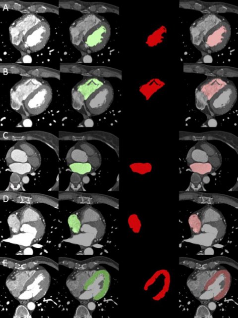
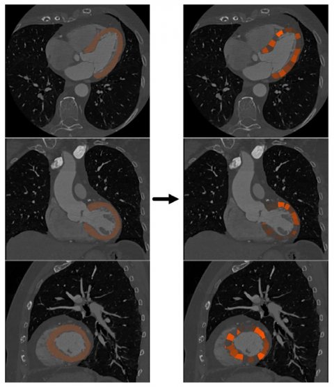
Tim Leiner, Chair of Cardiovascular Imaging in the Department of Radiology and Nuclear Medicine at the Utrecht University Medical Center in The Netherlands, highlighted how to apply AI in CMR and CT. With a role in automated identification of anatomical structures from CCTA, he pointed to AI being used to calculate the volumes of the chambers and LV mass from images. “This information is readily extractable and neural networks help you to do this in clinical routine and integrate the information into a report,” he continued. He also discussed automated identification of cardiac structures from non-contrast CT and deep learning for estimating the presence of flow-limiting coronary stenosis.
Professor Leiner detailed the role of AI acquisition and reconstruction of images, prognosis and prediction, such as a patient’s survival prediction based on right ventricular motion and how radiomics improves prediction of coronary events. “Machine learning and deep learning technique are exquisitely suited to derivation of prognosis and the key concept here is that images that are made for diagnostic purposes also contain prognostic information,” he told delegates. “That information may not be immediately relevant for clinical decision making today but it has information about what will happen to the patient in the future. This could, for instance, be used to adjust intensity of therapy or to direct expensive therapies to those most in need.”

He pointed to examples of expensive therapies for pulmonary hypertension – a condition with poor prognosis – to illustrate this, with new therapies costing up to $45,000 per patient a year. “In an increasingly cost-conscious environment you want to give this to people who need it most,” said Professor Leiner.
AI facilitates the use of neural networks to try to predict outcomes of patients to help in that decision making for who receives expensive drugs. It also helps highlight information that is difficult to extract visually from just looking at the images. He noted that there are challenges as well as opportunities in bringing machine learning to the clinic, as well as ethical considerations, but there are “great opportunities for clinically meaningful work.” “The entire imaging pipeline can be improved…and there are still some issues, but the future is very bright,” said Professor Leiner.
Piotr Slomka, Professor of Medicine and Cardiology in the Division of Artificial Intelligence in Medicine, in the Cedars-Sinai Medical Centre in Los Angeles, shifted the focus to applying AI in (hybrid) nuclear cardiology.
Stressing that data is key to developing good AI systems, he discussed deep learning for diagnosis, how AI can predict early revascularisation from nuclear cardiology, be used for automated coronary calcium scoring, epicardial adipose tissue quantification and forecasting long-term risk of major cardiovascular events. He said: “AI can assist in mental integration of multiple quantitative and clinical parameters and provide more accurate risk estimates after the imaging test, while deep learning can provide diagnosis estimate.”
Profiles:
Steffen Petersen is a Professor of Cardiovascular Medicine at the William Harvey Research Institute, Queen Mary University of London and a Consultant Cardiologist and Clinical Director for Research at Barts Heart Centre, Barts Health NHS Trust. He is also the Cardiovascular Programme Director of UCLPartners Academic Medical Centre and President-Elect of the European Association of Cardiovascular Imaging (EACVI).
Tim Leiner is Professor of Radiology and Chair of Cardiovascular Imaging at Utrecht University Medical Center, Utrecht, in The Netherlands. His research centres on the development and implementation of new MR and CT techniques with a focus on cardiovascular imaging. He is currently President of the International Society for Magnetic Resonance in Medicine (ISMRM).
Piotr Slomka is the director of Innovation in Imaging and professor of medicine and cardiology, Division of Artificial Intelligence in Medicine at Cedars-Sinai. Current research in the Slomka Laboratory focuses on developing innovative methods for fully automated analysis of nuclear cardiology data using novel algorithms and machine learning techniques.
28.08.2021


