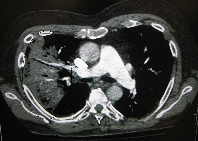
Credit: James Heilman, MD. Wikimedia Commons/CC By-SA 3.0
News • Survival of patients
Unsupervised AI predicts the progression of COVID-19
Unsupervised deep learning breaks new ground by predicting the progression of COVID-19 and survival of patients directly from their chest CT images.
Fast and accurate clinical assessment of the disease progression and mortality is vital for the management of COVID-19 patients. Although several predictors have been proposed, they have been limited to subjective assessment, semi-automated schemes, or supervised deep learning approaches. Such predictors are subjective or require laborious annotation of training cases.
In a multi-center study that was published in Medical Image Analysis, a research team lead by Hiroyuki Yoshida, Ph.D., director of the 3D Imaging Research at Massachusetts General Hospital (MGH), showed that unsupervised deep learning based on computed tomography can provide a significantly higher prognostic performance than established laboratory tests and existing image-based visual and quantitative survival predictors. The model can predict, for each patient, the time when COVID-19 progresses and thus the time when the patient is admitted to an intensive care unit or when the patient is diseased, something that other image-based prediction models cannot do. The time information calculated by the model also enables stratification of the patients into low- and high-risk groups by a wider margin than what is possible with other predictors.
"Our results show that the prediction performance of the unsupervised AI model was significantly higher and the prediction error significantly lower than those of the previously established reference predictors," says Yoshida. "The use of unsupervised AI as an integral part of the survival prediction model makes it possible to perform prognostic predictions directly from the original CT images of patients at a higher accuracy than what was previously possible in quantitative imaging."
In a companion study that was published recently in Nature, the team had already shown that supervised AI can be used to predict the survival of COVID-19 patients from their chest CT images. However, the new unsupervised AI model breaks new ground by avoiding the technical limitations and the laborious annotation efforts of the previous predictors, because the use of a generative adversarial network makes it possible to train a complete end-to-end survival analysis model directly from the images. "It is a much more precise and highly advanced AI technology," Yoshida explains.
Although the study was limited to COVID-19 patients, the team believes that the model can be generalized to other diseases as well. "Issues such as Long COVID, the Delta variant, or generalization of the model to other diseases manifested in medical images are promising applications of this unsupervised AI model," says Yoshida.
Source: Massachusetts General Hospital
01.09.2021


