Credit: Radiological Society of North America
News • Imaging AI
Photon-counting CT improves myeloma bone disease detection
New CT technology paired with artificial intelligence (AI)-based noise reduction offers superior detection of bone disease associated with multiple myeloma at lower radiation doses than conventional CT.
The photon-counting detector CT debuted in the clinic in 2021 after decades of development. By directly converting individual X-ray photons into an electric signal, photon-counting detector CT can decrease the detector pixel size and improve the image's spatial resolution.
"Additionally, photon-counting CT has demonstrated much better dose efficiency than standard CT, which allows for acquisition of ultra-high-resolution images of large areas of the body," said study lead author Francis Baffour, M.D., diagnostic radiologist at the Mayo Clinic in Rochester, Minnesota.
This potential for improved image quality in whole-body low-dose scans inspired Dr. Baffour and colleagues to study the technology in people with multiple myeloma, a disease that forms in a type of white blood cell found in the bone marrow called a plasma cell. Bone disease characterized by areas of bone destruction known as lytic lesions is found in approximately 80% of multiple myeloma patients.
The International Myeloma Working Group recommends low-dose, whole-body CT to evaluate associated bone disease. Much less is known about photon-counting detector CT is this setting.
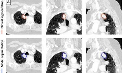
Dr. Baffour and colleagues compared photon-counting detector CT with conventional low-dose, whole-body CT in 27 multiple myeloma patients, median age 68 years. The patients underwent whole-body scans with both types of CT and two radiologists compared the images.
"We felt this was a prime example to showcase the ultra-high-resolution of photon-counting CT at low scanning doses," Dr. Baffour said.
Credit: Radiological Society of North America
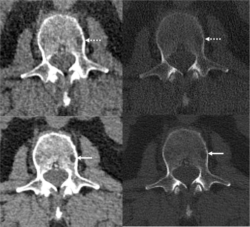
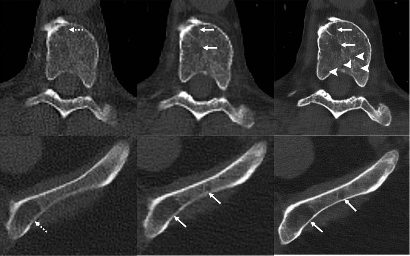
The researchers also applied a deep learning AI technique developed at Mayo Clinic's CT Clinical Innovation Center to reduce the noise in the very sharp photon-counting images. CT noise refers to an unwanted change in pixel values in the image, often loosely defined as the grainy appearance on cross-sectional imaging. The photon-counting detector CT with deep learning noise reduction demonstrated improvement in visualization and detected more lesions relative to conventional CT.
"We were excited to see that not only were we able to detect these features of multiple myeloma disease activity more clearly on the photon-counting scanner," Dr. Baffour said, "with deep learning denoising techniques that allowed us to generate thinner image slices, we were able to detect more lesions than on the standard CT.
The researchers hope to conduct follow-up studies on patients with multiple myeloma precursor states to see if photon-counting detector CT finds bone lesions that would upstage these patients to active multiple myeloma.
"Our excitement as scientists and radiologists in these results stems from our realization that this scanner could make a difference in the staging of disease, potentially impact therapy choice, and ultimately, patient outcomes."
They also want to look at photon-counting detector CT in other instances in which low-dose protocols are beneficial, for instance, in pediatric or pregnant patients or screening applications.
"Already there are ongoing studies to determine how low we can go with scanning doses while still obtaining diagnostic CT images," Dr. Baffour said. "So, there is much on the horizon and so much potential for photon-counting detector CT in clinical care."
The study was published in Radiology.
Source: Radiological Society of North America
07.09.2022