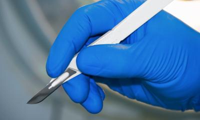
Image courtesy of Dr. L. Eberlin
News • Tissue identification
Mass spectrometry pen put to good use in thyroid surgery
Surgery of the thyroid and parathyroid glands is most challenging, even to expert surgeons. These relatively small structures located in the neck are in contact with each other and share certain features, including color and tactile feel, making it difficult to visually identify them.
“In procedures to remove the thyroid, for example, inadvertent parathyroid removal occurs in up to 25% of cases. When removing parathyroid glands, a common cause of unsuccessful procedures is the failure to localize and resect the diseased parathyroid tissue, as thyroid nodules and lymph nodes can be mistakenly identified as parathyroid tissue,” said co-corresponding author Dr. James Suliburk, associate professor of surgery and member of the Dan L Duncan Comprehensive Cancer Center at Baylor College of Medicine. “There is a pressing need to introduce innovative methods to preserve healthy tissue and ensure resection of only the intended tissue.”
The thyroid is shaped like a butterfly and sits in front of the windpipe or trachea. The parathyroid glands are four tiny structures located just to the side and behind the thyroid gland. Healthy parathyroid glands are each the size of two grains of rice and a slight shade different color from the yellow fat around them; sometimes they can be concealed by other nearby tissues. If the parathyroid glands become overactive, a surgeon may recommend surgical removal of one or more of the glands, the only cure for primary hyperparathyroidism. Likewise, in surgeries to remove a diseased thyroid, surgeons want to leave the parathyroids untouched.
In this study published in the journal JAMA Surgery, Drs. Livia S. Eberlin, Suliburk and their colleagues evaluated the performance and clinical applicability of a handheld probe called the MasSpec Pen, a technology for discriminating thyroid, parathyroid and lymph node tissues during surgery.
Recommended article

Article • Mass spectrometry goes handheld
A pen to pin down the fringes of cancer
Mass spectrometry – a powerful tool for analysing the molecular composition of a tissue sample – is invaluable during cancer surgery. However, mass spectrometers are complex and unwieldy, and certainly a poor fit for an operating room (OR). To create a bridge between the lab and OR, Professor Livia S Eberlin, from Baylor College of Medicine, has developed a very special ‘pen’.
“In this project, we tested the MasSpec Pen extensively during thyroid and parathyroid surgery,” said Eberlin, Translational Research and Innovations Endowed Chair and associate professor of surgery at Baylor. She also is co-corresponding author of the work. “The MasSpec Pen primarily detects small molecules – metabolites (products of cell metabolism) and lipids made by cells. The pattern of metabolites and lipids is very characteristic of each tissue. Even though many of the molecules made by different tissues are similar, their abundance differs depending on the tissue type, and this allows us to distinguish one tissue from another.”
“It’s similar to when you make coffee,” Eberlin said. “You run water through the coffee grinds to extract molecules from it.”
Then, a tubing carries the water droplet to a mass spectrometer, an instrument that in real time reads the molecular composition of the droplet, which reveals the type of tissue it came from. “The procedure causes no damage to the tissues being analyzed,” Eberlin said.
“In the operating room, the MasSpec Pen can help surgeons identify the tissue that they are considering to resect before actually resecting it, to ensure that it was the right tissue and no tissue was removed unnecessarily,” Suliburk said. “The pen integrates naturally into the surgical workflow. We sterilize it just as our surgical instruments and connect it to the mass spectrometer just like we connect other instruments in the operating room. It is very intuitive to use and has immense potential for saving time during surgery.”
To evaluate the accuracy of this new technology, the researchers compared the tissue identification provided by the MasSpec Pen with the golden standard of tissue identification, pathology analysis. A pathologist is a medical professional specialized in identifying tissue samples under the microscope. “The MasSpec Pen showed high performance for discriminating thyroid, parathyroid and lymph node tissues during surgery, with more than 90% accuracy,” Eberlin said.
“Also, we had the results of the MasSpec Pen analysis in about 20 seconds, whereas the processing of the sample for pathology analysis during surgery (called frozen section) can take up to an hour,” Suliburk said. “This adds time and cost to the procedure. Typically, the longer the operation, the higher the risk of complications. Using the pen benefits the patient by offering the power of real-time tissue identification as we operate. This work is a wonderful example of how live tissue sensing can improve patient care. The goal is to expand its use to a variety of different projects where live tissue identification could be very powerful. The MasSpec Pen could be applied to surgeries of other organs, such as lungs or pancreas,” Suliburk said. “We think this can really revolutionize how we do surgery.”
Other contributors to this work include Rachel J. DeHoog, Mary E. King, Michael F. Keating, Jialing Zhang, Marta Sans, Clara L. Feider, Kyana Y. Garza, Alena Bensussan, Anna Krieger, John Q. Lin, Sunil Badal, Elizabeth Alore, Christopher Pirko, Kirtan Brahmbhatt, Wendong Yu and Raymon Grogan. The authors are affiliated with Baylor College of Medicine and/or the University of Texas at Austin.
This work was supported by the National Cancer Institute (R33CA229068-01), the Gordon and Betty Moore Foundation (GBMF8049) and the Welch Foundation (F-1895).
Source: Baylor College of Medicine
31.08.2023