Article • Exploring imaging advances and their impact on radiographers
‘The future of breast MRI is bright’
Breast MRI has emerged as a powerful diagnostic tool, particularly for women with dense breast tissue where traditional mammography faces limitations. In her presentation at ECR 2025, radiographer Hanna Kalliomäki highlighted several technological advances transforming breast cancer detection and diagnosis. From time-saving abbreviated protocols and AI-assisted analysis to contrast-free diffusion techniques and molecular imaging, these techniques are reshaping how radiographers approach breast cancer screening and diagnosis.
Article: Wolfgang Behrends

The expert from the Päijät-Häme Wellbeing County in Lahti, Finland, explained that breast MRI offers exceptional sensitivity for screening high-risk women and evaluating tumour extent, treatment response, and implant integrity.1 While mammography remains an effective screening tool, its sensitivity decreases significantly in dense breast tissue, due to the masking effect of fibroglandular tissue. ‘Since almost half of the screening population has dense breasts, many of these patients require additional MRI breast imaging,’ she pointed out.
AB-MRI: a game changer in patient throughput and comfort
One of the most significant developments is Abbreviated Breast MRI (AB-MRI), which reduces scan times from 30-60 minutes to just ten minutes without compromising diagnostic accuracy.2 ‘This allows MRI to be used as a screening tool for a broader population, not just those at high risk for breast cancer,’ Kalliomäki said, adding that ‘AB-MRI is a game changer in terms of patient throughput and comfort. Shorter scan times reduce patient anxiety, minimize motion artifacts, and allow imaging more patients without compromising diagnostic accuracy.’
For radiographers, mastering AB-MRI protocols and optimizing scan parameters for both speed and accuracy represents an important skill development opportunity. ‘You'll also need to explain the benefits to patients who may be apprehensive about MRI due to the traditionally long durations,’ she pointed out.
AI-augmented workflows
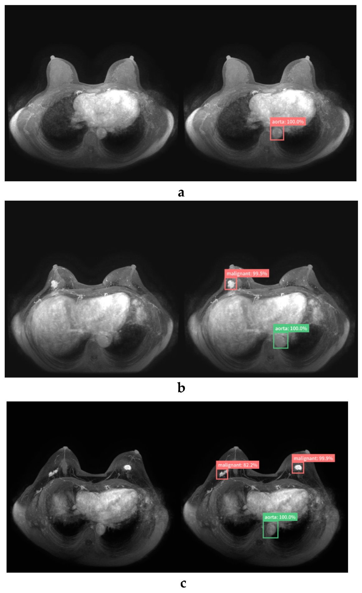
Image source: Adachi M, Fujioka T, Mori M et al., Diagnostics 2020 (CC BY 4.0)
Kalliomäki further explored the benefits of artificial intelligence (AI) in image data analysis and lesion classification.3,4 Able to detect subtle abnormalities that might be missed by a human observer, these systems help reduce both false positives – which lead to unnecessary biopsies and patient anxiety – and false negatives – missed cancers.
Despite this, the expert reassured her audience that ‘AI won't replace radiographers but will augment your work’. Radiographers will continue to play vital roles in patient positioning and ensuring high-quality scans – prerequisites for effective AI analysis. Still, understanding how AI tools integrate with imaging workflows will be essential for radiographers moving forward, she stressed.
Contrast-free imaging and personalized profiles
Advances also include promising new imaging techniques, such as Diffusion-Weighted Imaging (DWI). Kalliomäki explained how DWI is able to distinguish between benign and malignant tissues without contrast agents by highlighting areas of abnormal water molecule movement. ‘This technique enhances the safety of breast MRI for a wider range of patients, especially for patients with contrast allergies or kidney complications,’ she said. Since DWI is sensitive to motion, proper patient positioning becomes even more critical. Radiographers must understand DWI settings and how to adjust them for different patients to obtain high-quality diagnostic images.5
Another approach that is becoming increasingly common in breast imaging is molecular MRI, which uses targeted contrast agents that bind to specific cancer cells or biomarkers like HER2 receptors. ‘This approach visualizes tumour characteristics at the molecular level, offering a personalized approach to imaging,’ Kalliomäki pointed out. ‘This enhances detection and diagnosis, tailors imaging to patients' profiles, and support targeted therapies like those for HER2-positive breast cancer, potentially improving survival rates and reducing overtreatment.’
The new innovations are poised to reshape how we screen for and diagnose breast cancer
Hanna Kalliomäki
The expert advised her colleagues to familiarize themselves with these new, specialized agents, as radiographers will need to develop expertise in their safe administration and optimal use within imaging protocols. ‘This means you will be part of a team that tailors imaging to each patient’s cancer type. Accurate timing and precise contrast administration will be key as these agents are designed to highlight specific tumour characteristics.’
Kalliomäki also highlighted two promising new techniques; breast MRI spectroscopy and synthetic correlated diffusion imaging (CDI). While the former analyses the chemical composition of breast tissue for specific metabolites elevated in tumours,6 the latter synthesizes multiple DWI images from raw data.7 ‘By offering biochemical insights beyond structural imaging, spectroscopy adds another dimension to comprehensive breast assessment,’ she said.
‘Imaging is teamwork’
‘The future of breast MRI is bright,’ Kalliomäki concluded, pointing out the AI-driven precision diagnostics poised to revitalize the field. The advent of non-contrast imaging techniques and whole breast MRI screening for dense breasts was also welcomed by the expert, who only expressed some concern over implementation challenges due to limited MRI availability.
Kalliomäki concluded her presentation with an important reminder: ‘The new innovations are poised to reshape how we screen for and diagnose breast cancer.8,9 Radiographers are integral for the successful implementation of these advances in breast MRI technologies. The expertise of radiographers and radiologists together ensures imaging protocols are optimized, leading to improved diagnostic accuracy and enhancing patient care. Imaging is teamwork.’
References:
- Mann RM, Balleyguier C, Baltzer PA et al.: Breast MRI: EUSOBI recommendations for women's information; European Radiology 2015; https://doi.org/10.1007/s00330-015-3807-z
- Sardanelli F, Magni V, Rossini G, Kilburn-Toppin F, Healy NA, Gilbert FJ: The paradox of MRI for breast cancer screening: high-risk and dense breasts—available evidence and current practice; Insights into Imaging 2024; https://doi.org/10.1186/s13244-024-01653-4
- Adachi M, Fujioka T, Mori M et al.: Detection and Diagnosis of Breast Cancer Using Artificial Intelligence Based assessment of Maximum Intensity Projection Dynamic Contrast-Enhanced Magnetic Resonance Images; Diagnostics 2020; https://doi.org/10.3390/diagnostics10050330
- Chen Y, Shao X, Shi K, Rominger A, Caobelli F: AI in Breast Cancer Imaging: An Update and Future Trends; Seminars in Nuclear Medicine 2025; https://doi.org/10.1053/j.semnuclmed.2025.01.008
- Baltzer P, Mann RM, Iima M et al.: Diffusion-weighted imaging of the breast—a consensus and mission statement from the EUSOBI International Breast Diffusion-Weighted Imaging working group; European Radiology 2020; https://doi.org/10.1007/s00330-019-06510-3
- Bitencourt A, Sevilimedu V, Morris EA, Pinker K, Thakur SB: Fat Composition Measured by Proton Spectroscopy: A Breast Cancer Tumor Marker?; Diagnostics 2021; https://doi.org/10.3390/diagnostics11030564
- Tai CA, Gunraj H, Hodzic N et al.: Enhancing Clinical Support for Breast Cancer with Deep Learning Models Using Synthetic Correlated Diffusion Imaging. In: Wu S, Shabestari B, Xing L (eds): Applications of Medical Artificial Intelligence; Applications of Medical Artificial Intelligence; https://doi.org/10.1007/978-3-031-47076-9_9
- Galati F, Trimboli RM, Pediconi F: Special Issue “Advances in Breast MRI”; Diagnostics 2021; https://doi.org/10.3390/diagnostics11122297
- Bougias H, Stogiannos N: Breast MRI: Where are we currently standing?; Journal of Medical Imaging and Radiation Sciences 2022; https://doi.org/10.1016/j.jmir.2022.03.072
22.04.2025