Image source: Stephan Wiegand/Hochschulmedizin Dresden
News • "In-beam MRI"
Radiation therapy: proton trajectory made visible
The aim of proton radiation therapy in fighting cancer is to kill as many tumor cells as possible while also protecting the surrounding healthy tissue. As there is yet no direct method for mapping the beam range during dose delivery, physicians for example at the University Hospital Dresden work with safety margins around the tumor that affect the conformity of dose distribution and reduce accurate targeting.
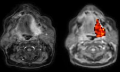
Dresden scientists led by Prof. Aswin L. Hoffmann from the National Center for Radiation Research in Oncology – OncoRay, which is operated by the Helmholtz-Zentrum Dresden-Rossendorf (HZDR), among others, have succeeded in visualizing the proton beam’s trajectory in a fluid-filled phantom using an “in-beam MRI” prototype. By utilizing this method they revealed the proton beam’s range during irradiation. The team published the results in the journal Proceedings of the National Academy of Science.
Unlike photons, protons possess an important advantage: they have a defined range – that is, a point at which they release their maximum energy. In proton radiation therapy, this property makes it possible to stop the radiation within the tumor tissue and apply a high dose of radiation in that area, while at the same time greatly reducing the dose delivered to the surrounding healthy tissue. Proton radiation therapy is therefore mainly used to treat children, but also adults with tumors located near normal tissue that is highly sensitive to radiation. To control dose delivery, a direct method is required that measures and images the beam’s range in relation to the patient’s anatomy during irradiation. Because such a method is yet unavailable, safety margins have so far been used around the tumor tissue, which leads to irradiation of normal tissue and limits the maximum possible dose in the tumor.
Image source: Stephan Wiegand/Hochschulmedizin Dresden
The group led by Hoffmann has been researching the technical integration of magnetic resonance imaging (MRI) and proton therapy since 2016. Using an “in-beam MRI” prototype, Hoffman and his group have succeeded for the first time in the world in visualizing the proton beam in a fluid-filled phantom and in using this method to reveal the proton beam’s range during radiation. “The result of our work could significantly alter the quality assurance of proton therapy. Previously, measurements were often made indirectly, but now proton beam imaging can occur directly during dose application,” Hoffman explains. “My dream is to be able to apply this method in the future for monitoring patient treatments.”
The study demonstrated the feasibility of proton beam visualization in liquid media. As predicted, the MRI images obtained during irradiation showed that the penetration depth increased with increasing proton energy, and therefore the strength of the MRI signal also increased with the increasing proton current. “This result is an important step in image-guided proton therapy,” says Prof. Mechthild Krause, director of OncoRay. “Real-time MRI imaging has already made its way into conventional photon radiotherapy. Professor Hoffmann and his team are working on a prototype for a new irradiation device that should also establish real-time MRI imaging in proton therapy.”
The results from Hoffmann’s group provide hope that a new dimension in cancer patient treatment will be possible. Currently, a new large-scale MRI system is being installed in the OncoRay building. This will make it possible for the first time to perform proton irradiation and real-time MRI simultaneously, while it will also provide the ability to vary direction and strength of the magnetic field relative to the patient. This could enable a more precise use of proton therapy in a few years for mobile tumors in that it will even better preserve the healthy tissue and will irradiate the tumor tissue with a higher dosage.
Source: Helmholtz-Zentrum Dresden-Rossendorf
24.06.2023