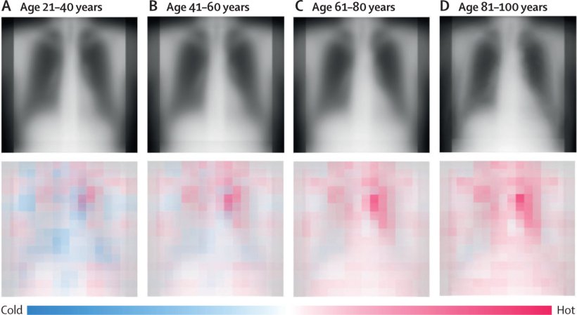
Image source: Mitsuyama et al., Lancet Healthy Longevity 2023 (CC BY 4.0)
News • Radiograph evaluation
AI uses chest X-rays to determine "true" age of a patient
What if “looking your age” refers not to your face, but to your chest? Osaka Metropolitan University scientists have developed an advanced artificial intelligence (AI) model that utilizes chest radiographs to accurately estimate a patient’s chronological age.
More importantly, when there is a disparity, it can signal a correlation with chronic disease. These findings mark a leap in medical imaging, paving the way for improved early disease detection and intervention. The results are published in The Lancet Healthy Longevity.
The research team, led by graduate student Yasuhito Mitsuyama and Dr. Daiju Ueda from the Department of Diagnostic and Interventional Radiology at the Graduate School of Medicine, Osaka Metropolitan University, first constructed a deep learning-based AI model to estimate age from chest radiographs of healthy individuals. They then applied the model to radiographs of patients with known diseases to analyze the relationship between AI-estimated age and each disease. Given that AI trained on a single dataset is prone to overfitting, the researchers collected data from multiple institutions.
Our results suggest that chest radiography-based apparent age may accurately reflect health conditions beyond chronological age
Yasuhito Mitsuyama
For the development, training, internal and external testing of the AI model for age estimation, a total of 67,099 chest radiographs were obtained between 2008 and 2021 from 36,051 healthy individuals who underwent health check-ups at three facilities. The developed model showed a correlation coefficient of 0.95 between the AI-estimated age and chronological age. Generally, a correlation coefficient of 0.9 or higher is considered to be very strong.
To validate the usefulness of AI-estimated age using chest radiographs as a biomarker, an additional 34,197 chest radiographs were compiled from 34,197 patients with known diseases from two other institutions. The results revealed that the difference between AI-estimated age and the patient’s chronological age was positively correlated with a variety of chronic diseases, such as hypertension, hyperuricemia, and chronic obstructive pulmonary disease (COPD). In other words, the higher the AI-estimated age compared to the chronological age, the more likely individuals were to have these diseases.
“Chronological age is one of the most critical factors in medicine,” stated Mr. Mitsuyama. “Our results suggest that chest radiography-based apparent age may accurately reflect health conditions beyond chronological age. We aim to further develop this research and apply it to estimate the severity of chronic diseases, to predict life expectancy, and to forecast possible surgical complications.”
Source: Osaka Metropolitan University
26.08.2023