
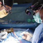
News • Microscopy
New microscopy method maps lipids without labels
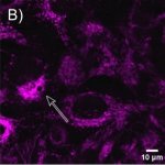
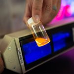
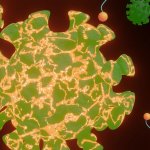
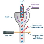
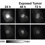
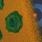
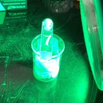
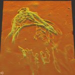
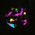
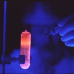
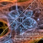
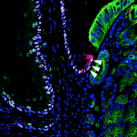
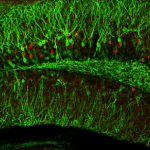
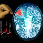
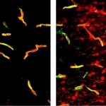
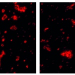
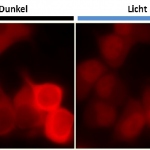
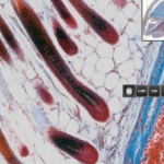
A new microscopy method reads lipids' natural spectral fingerprints using mid-infrared light and ultrasound – no fluorescent labels needed, less stress for living cells.


A new microscopy method reads lipids' natural spectral fingerprints using mid-infrared light and ultrasound – no fluorescent labels needed, less stress for living cells.
A new analytical method could improve how cancer treatments are designed – by allowing scientists to track, for the first time, exactly where inside a living cell a drug accumulates.
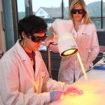
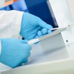
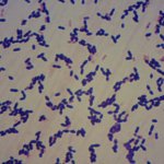
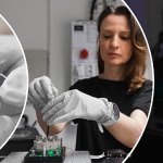
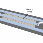
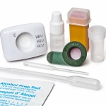
A research team is leading the development of a sensor that paves the way for the rapid, selective and cost-effective detection of active tuberculosis infection.

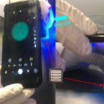
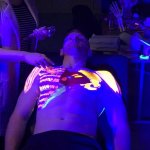
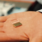
A compact LED module pairs UV-A, green and IR light with a matched photodiode to detect AGE autofluorescence — bringing biochemical risk screening to wearables and point of care testing.
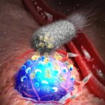
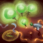
A research team led by the University of Waterloo is developing a novel tool to treat cancer by engineering hungry bacteria to literally eat tumours from the inside out.
Highlights:Shimadzu is offering a wide range of solutions in liquid chromatography starting from standard HPLC systems to high-end UHPLC systems including compact configurations. Available with several options for columns switching, pre-concentration, online SPE, etc, the systems are also well recognized for coupling with highly sensitive detectors like fluorescence, radio-activity,…
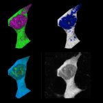
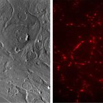
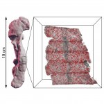
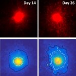
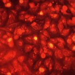
A new type of 3D imaging reveals how amyloid β (Aβ) deposits spread along blood vessels in the human brain. This could lead to targeted therapies for Alzheimer's disease and CAA.
Scientists have developed a new method to track the build-up of amyloid plaques – a key characteristic of Alzheimer’s disease – in real time – an important step forward towards new treatments.
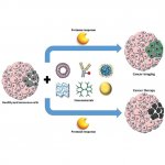
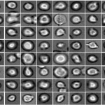
Scientists created precise replicas of Candida sugar coats to understand immune responses, enabling fast bedside testing that could replace slow lab cultures.
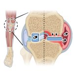
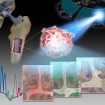
A squishy new ‘artificial cartilage’ material could improve arthritis treatments by releasing anti-inflammatory drugs in response to a flare-up.
An advanced imaging method that uses the natural glow of tissues could help detect subtle differences in the tissue’s biochemistry, offering a way to earlier detect colorectal cancer via endoscopy.
Tracer molecules serve to provide visual guidance in nuclear medicine, but a new molecule could give surgeons additional audio cues to help them locate prostate cancer tumors and metastases.

Tumor vesicles may serve as early indicators of cancer and help monitor the effectiveness of treatments. Now, researchers discovered a new way to detect these vesicles.
How cancer cells survive the journey through the body to form metastases is still poorly understood. New insights into how cells survive intense physical stress could pave the way for new treatments.
A new, bacteria-based contrast agent illuminates tumors like a neon sign during surgery, enabling more precise resection and reducing the risk of recurrence.
Through quantitative real-time monitoring of cell spatiotemporal dynamics, or cell changes over time, electrical impedance spectroscopy (EIS) could be used to predict cancer growth.
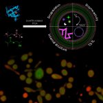
In a new study, researchers have developed a new fluorescent probe to visualise signaling dynamics in moving cancer cells, to uncover a new therapeutic possibility for limiting breast cancer spread.
Researchers used state-of-the-art 3D bioprinting to replicate the complex structure and environment of spinal discs. Their insights hold promise for understanding back pain and disc degeneration.
A new approach enables the detection of even trace amounts of cancer cells. The method leverages SERS for signal amplification, eliminating the need for fluorescent dyes.

World TB Day raises awareness about tuberculosis and commemorates the discovery of the source bacterium M. tuberculosis. More than a century later, scientists still refine anti-TB strategies.
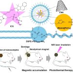
Japanese researchers have developed magnetic nanoparticles that can be directed to tumors using a magnet and then heated with a laser to destroy cancer cells while sparing healthy tissue.
US researchers have developed a comprehensive deep learning AI model designed to more accurately identify and classify cells in high-content tissue images.
Molecular pathology should become centralised in fewer labs to improve efficiencies and affordability, according to leading European experts.

Multiplex imaging can play a critical role in unravelling the tumour microenvironment. The potential and benefits of the emerging approach – a way to extract information from human tissue samples by visualising many more biomarkers than traditional microscopy – was highlighted in presentations during the 36th European Congress of Pathology in Florence, Italy. Speakers also discussed novel…

The year: 2034. Breast cancer patients benefit from perfectly personalised diagnostics and therapies. The tedium of follow-up treatments is a thing of the past, thanks to AI, augmented reality and robotics. Just a tale from the realm of science fiction, or could this soon be clinical reality? At the annual meeting of the German Senologic Society, Prof Dr Marc Thill from the Agaplesion Markus…
New insights into metastasis: Scientists created a 3D-printed model to mimic the specific conditions that spur the spread of cancer cells. This could help discover new screening and treatment options.
Before administering certain drugs, doctors check a patient’s kidney function by testing their blood urea nitrogen and creatinine levels. New research shows that gold nanoparticles might give more accurate results.
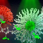
Researchers propose the use of molecular ‘cages’ (made of pseudopeptides) to selectively eliminate cancer cells in acidic microenvironments. This could help reduce side effects from chemotherapy.
Researchers discovered islands of highly potent immune cells in the vicinity of glioblastomas. This may open up prospects for new therapies for these aggressive brain tumours.
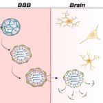
Researchers were able to package fluorescent sensors for passage across the blood-brain barrier (BBB) in mice. This holds promise to advance Alzheimer’s disease diagnosis and treatment.
Researchers have developed a fluorescence assay for viral integrity (or FAIRY, for short), to quickly determine the effectiveness of countermeasures against a given virus.

Endoscopy is pivotal in diagnosing and managing ulcerative colitis. Recent technology advances allow for early cancer detection, precise disease assessment and targeted biopsies, improving diagnosis and monitoring. The following article takes a look at the latest advancements.
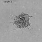
After a stroke or heart attack, the risk of infection is increased – however, why this happens was unknown. Now, researchers found a previously unknown cause – and a therapeutic approach.
Researchers have discovered how the mechanical properties of tumours - their softness, for example - can prime cancer cells to better survive their spread to other organs.

Digital pathology can be used to great effect in pharmaceutical research: it can accelerate analyses, give deeper insights into cellular mechanisms, and enable better understanding of their role into clinical development. This potentially offers clearer predictions on how patients may respond to treatment and lead to personalized therapies.
A research team created "laboratory testicles" that may significantly advance understanding of the mechanisms involved in sex determination and provide solutions for male infertility.

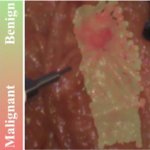
Breast surgery is a traumatic experience for a woman, no matter whether breast-conservation surgery (BCS) or a mastectomy. Trauma levels are greatly enhanced, if pathological evaluation findings of an excised breast tumour following a lumpectomy suggest that additional cancer may still be in the margins, and a second surgical procedure is required. A new system with the ability to accurately…
A new soldering technique developed by Empa researchers is expected to prevent wound healing disorders and life-threatening complications from leaking sutures.
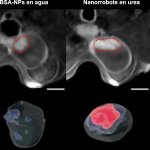
New research demonstrates how tiny nanomachines could greatly reduce bladder cancer by precisely targeting the tumour and attacking it with a radioisotope carried on their surface.
Researchers have developed a simple, yet efficient way to create cell-like synthetic structures. These artificial cells could be used for a range of medical purposes – for example, in drug delivery.

Breaking & entering – on a minuscule scale: US researchers have shown how some pathogens like Toxoplasma can enter cells using physical force to cause an infection.

A new deep-learning approach to AMR testing has been shown to detect antimicrobial susceptibility within as little as 30 minutes - significantly faster than current gold-standard approaches.
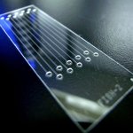
Faster, more accurate and cost-effective testing: Experts outline the beginnings and evolution of “lab-on-a-chip” technology, and its benefits for advanced and next-gen operational platforms.
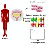
In a new study, researchers from Fudan University have developed a novel urine-based prognostic model that promises to transform the management and treatment of bladder cancer.

Inspired by the enhanced visual system of butterflies, researchers have developed an imaging sensor to “see” into the UV range for differentiating between cancer and normal cells.

Würzburg resarchers have created a new pseudovirus design that allows tracking of penetration of viruses into cells.
A team of researchers has developed a visualisation tool that combines high-speed cameras and fluorescent injection to distinguish tumour tissue from normal tissue across cancer types.
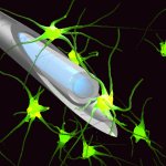
Half as thick as a human hair: Researchers at TU Munich have developed the world's first microrobot capable of navigating within groups of cells and stimulating individual cells.
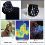
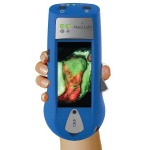
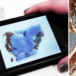
Scientists have developed a device that works with a smartphone or tablet to capture medical images which can identify infected wounds through thermal and fluorescence imaging.

Under the impulse of the European Commission, the in vitro diagnostic industry is developing emerging technologies to implement sustainable practices in medical laboratories. As sustainability has been a growing priority of the European Union (EU) in the last decade, ‘the medical technology sector, particularly the IVD sector, must comply with European legislation in this field like all other…
Plastics are a part of everyday life, and an increasingly concerning factor of global environmental pollution. They also have infiltrated our bodies as microparticles (MPs) and nanoparticles (NPs), found even in placentas supporting foetal life. And they are in our blood. Now, researchers in Spain have developed a new method to detect and measure nanoparticles in human peripheral blood that is…
Researchers at Harvard Medical School developed a new tool that promises to improve the way pathologists see and evaluate a tumor by providing detailed clues about the cancer.

Lyme disease, the most common tick-transmitted bacterial infection in the world, is challenging to diagnose. Initial early-stage symptoms may include skin rash, fever, headache, fatigue, swollen lymph nodes, and/or body and joint aches. However, these symptoms are also associated with many other diseases and medical conditions.
Getting vaccines to people who need them isn't always easy. Many vaccines require cold storage, making it difficult to ship them to remote areas that don't have the necessary infrastructure.
A UK research team has developed a new technique that combines machine learning with short-wave infrared (SWIR) fluorescence imaging to detect precise boundaries of tumors.
A new real-time imaging technique that uses a type of infrared light has, for the first time, been used during surgery to differentiate between cancerous tumours and healthy tissue.
Hamamatsu Photonics announces the release of a new high-speed kinetic plate imager, the FDSS-GX, designed for kinetic cell-based assay development.

New research will bring together scientists from across the globe to accelerate fluorescence-guided surgery for bone cancer patients. The upcoming trial is focused on the dye indocyanine green (ICG).
Researchers tracked viruses passing through face masks and compared their failure on the filter layers of different mask types. This could accelerate the development of surfaces that can kill viruses.
Researchers have created a tool that maps how breast cancer grows in previously unseen detail, and highlights how the cells around the tumour may be the key to controlling the spread of disease.
Speed or accuracy? As far as Covid-19 tests go, this was the choice you had to make. In the future, this dilemma could be a thing of the past.

With the rise of syndrome-style infections, co-infections and the current antimicrobial resistance challenges, the need for multiplexed diagnostics is now more important than ever.
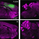
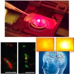
Treatment of central nervous system diseases and tumors is often hindered by the blood-brain barrier. A new method aims to overcome this obstacle using focused ultrasound intranasal delivery (FUSIN).
Researchers propose a new approach to identify cancerous tissue for surgical removal, based on real-time imaging of tissue oxygen concentration.
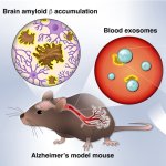
Researchers from Japan have developed a method to detect build-up of amyloid β in the brain, a characteristic of Alzheimer’s disease, from biomarkers in blood samples.
Researchers at Queen’s University Belfast have developed a new plastic film that can kill viruses that land on its surface with room light.
A new study could one day help health workers determine whether bacteria of the species Streptococcus pneumoniae, which cause meningitis, are resistant to antibiotics.

German researchers present a novel method for testing chemical agents that could help in the development of drugs against neurodegenerative diseases such as Alzheimer's.
The prototype device combines eRapid and SHERLOCK technologies into a single, postcard-sized system that can simultaneously detect the presence of both SARS-CoV-2 RNA and antibodies in a patient’s saliva.
The development of a simple blood test for glioblastomas could mean earlier diagnosis and more effective and personalised treatment options against the most common type of malignant brain cancer.
Research team led by Göttingen University combine two techniques to achieve isotropic super-resolution imaging.
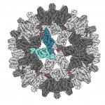
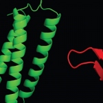
To develop new drugs, detailed knowledge about nature’s smallest biological building blocks is required. A new microscopy technique that allows proteins, DNA and other tiny biological particles to be studied in their natural state.
Researchers have developed a biodegradable gel that can help to improve the delivery of cells into the living heart and could form a new generation of treatments to repair heart attack damage.
Researchers have shown that aggregation of amyloid-beta, one of two key proteins implicated in Alzheimer’s disease, causes cells to overheat and ‘fry like eggs.’
An international team has for the first time demonstrated that nerve signals are exchanged between clogged up arteries and the brain.
Researchers have established an injectable hybrid inorganic nanoscaffold-templated stem cell assembly and applied it to the regeneration of critically-sized cartilage defects.
A chip-based infection model developed by Jena researchers enables live microscopic observation of damage to lung tissue caused by the invasive fungal infection aspergillosis.

Point-of-care testing (POCT) is a “win-win” scenario for patients and healthcare professionals in delivering care when and where it is needed, according to pathologist Adil Khan, MSc, PhD.
MediSCAPE, a high-speed 3D microscope, can see real-time cellular detail in living tissues to guide surgery, speed up tissue analyses, and improve treatments.
An ECRC research team has introduced CRISPR-Cas9 into human muscle stem cells for the first time using mRNA, thus discovering a method suitable for therapeutic applications.
Using a novel probe for functional magnetic resonance imaging, researchers have devised a way to monitor individual populations of neurons and reveal how they interact with each other.
A biocompatible ultrasound transducer chip could be a more effective way to harness the technology for biomedical applications.
Researchers in Zurich have developed a fluorescent orexin biosensor to observe on of the brain's signaling molecules "live" to gain insights into constant daytime sleepiness (narcolepsy).
Researchers have developed an inexpensive, non-toxic coating for almost any fabric that decreases the infectivity of the virus that causes COVID-19 by up to 90 per cent.
Heart cells from a patient with an inherited heart disease called arrhythmogenic cardiomyopathy do not contract correctly when grown in the laboratory, researchers from Osaka University have found.
A new biopsy tool will enable scientists and clinicians to simultaneously profile many biomarkers in cells and tissues.

Researchers have developed a diagnostic for Sars-CoV-2 that is capable of differentiating between Covid-19 and the garden-variety bug with fast turnaround.
Scientists at have designed a quantum sensor to detect SARS-CoV-2 faster, cheaper, and more accurate than the current gold-standard technique, PCR.
Ever since the Abbe diffraction limit of conventional microscopy has been surpassed, super-resolution techniques have been diving ever deeper into the most miniscule details of molecular structures. We spoke with Prof. Dominic Zerulla, whose company PEARlabs is developing an imaging technique that sets out to push the boundaries once more – by looking at in-vivo nano-scale processes in motion.
Test could measure patient immunity against multiple COVID-19 variants such as Omicron and Delta at once and inform which synthetic monoclonal antibody to use for treatments.
According to scientists at Rice University’s George R. Brown School of Engineering, discarded polystyrene broken down into microplastics provides a cozy home not only for microbes and chemical contaminants but also for the free-floating genetic materials that deliver to bacteria the gift of resistance.
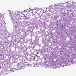
Professor Dina Tiniakos, from the National and Kapodistrian University of Athens, predicts that NASH (Non-alcohol related steatohepatitis) cases will soar worldwide by 2030, with 800,000 liver deaths, costing health economies billions of dollars.
Wearable technology has become an important part of medicine, from tracking vital signs to disease diagnosis. In surgery, wearable technologies can now assist, augment, and provide a means of patient assessment before, during and after surgical procedures. Wearable technologies are applied before the patient even reaches the operating room, for example in prehabilitation, i.e. pre-treatment…
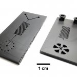
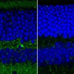
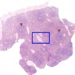
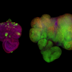
Researchers at Umeå University now demonstrate a method by which specific cell types in human organs can be studied with micrometer precision. The method can be used to reveal previously unrecognised alterations in the pancreas, but it can also be used to study other human organs and diseases.
Scientists use super-resolution microscopy to study previously undiscovered cellular worlds, revealing nanometer-scale details inside cells. This method revolutionized light microscopy and earned its inventors the 2014 Nobel Prize in Chemistry. In an international collaboration, AI researchers from Tübingen have now developed an algorithm that significantly accelerates this technology.
Nanocontainers can transport substances into cells where they can then take effect. This is the method used in, for example, the mRNA vaccines currently being employed against Covid-19 as well as certain cancer drugs. In research, similar transporters can also be used to deliver labelled substances into cells in order to study basic cellular functions. To take advantage of their full potential,…
A research team led by scientists in the labs of Jennifer Doudna, David Savage and Patrick Hsu at the University of California, Berkeley, is aiming to develop a rapid Covid-19 diagnostic test that is much faster and easier to deploy than qRT-PCR. It has now combined two different types of CRISPR enzymes to create an assay that can detect small amounts of viral RNA in less than an hour. Doudna…
Using lab-created tissue to heal or replace damaged organs is one of the great visions for the future of medicine. Synthetic materials could be suitable as scaffolding for tissue because, unlike natural tissues, they remain stable in the organism long enough for the body to form new natural structures. A fundamental requirement for functional tissue is that blood vessels must be able to grow in…
Researchers at RMIT University in Australia have developed smart wound dressings with built-in nanosensors that glow to alert patients when a wound is not healing properly. The multifunctional, antimicrobial dressings feature fluorescent sensors that glow brightly under UV light if infection starts to set in and can be used to monitor healing progress.
When viruses infect cells, changes in the cell nucleus occur, and these can be observed through fluorescence microscopy. Using fluoresence images from live cells, researchers at the University of Zurich have trained an artificial neural network to reliably recognize cells that are infected by adenoviruses or herpes viruses. The procedure also identifies severe acute infections at an early stage.
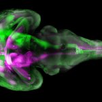
Researchers from ETH Zurich and University of Zurich have developed a new microscopy technique that lights up the brain with high resolution imagery. This allows neuroscientists to study brain functions and ailments more closely and non-invasively.
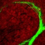
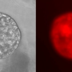
Before the huge potential of tiny nanocarriers for highly targeted drug delivery and environmental clean-up can be realized, scientists first need to be able to see them. Currently researchers have to rely on attaching fluorescent dyes or heavy metals to label parts of organic nanocarrier structures for investigation, often changing them in the process. A new technique using chemically-sensitive…
University of Birmingham scientists have developed a new microscopic imaging approach to take a closer look at 3D-printing for developing future patient implants, as well as improved disease modelling and drug screening. Additive manufacturing (3D printing) platforms create bioprinted structures by moving a special bioink, containing cells, biomolecules and materials, through a narrow tube, but…

Vigorous and rapid air exchanges might not always be a good thing when it comes to addressing levels of coronavirus particles in a multiroom building, according to a new modeling study. The study suggests that, in a multiroom building, rapid air exchanges can spread the virus rapidly from the source room into other rooms at high concentrations. Particle levels spike in adjacent rooms within 30…

Now Available: The Fluxergy Analyzer System is a testing platform designed for sample-to-answer point-of-care testing, enabling clinicians to cost-effectively conduct molecular in vitro diagnostic tests. The analyzer system is designed to deliver test results within one hour. This device is solely intended to be used by healthcare professionals.
What’s in a name? In the case of Asensus Surgical, Inc., previously known as TransEnterix, Inc., the recent rebranding doubles as a mission statement for the manufacturer of surgical robotics systems: The initial ‘A’ stands for artificial intelligence and augmented surgery, reflecting the company’s emphasis on new technologies designed to enhance the operator’s cognition (‘sensus’…
A research team of pharmacists at the University of Bonn has discovered two families of active substances that can block the replication of the SARS-CoV-2 coronavirus. The drug candidates are able to switch off the the key enzyme of the virus, the so-called main protease. The study is based on laboratory experiments. Extensive clinical trials are still required for their further development as…
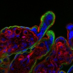
An improved high-tech fluorescence microscopy technique is allowing researchers to film cells inside the breast as never seen before. This new protocol provides detailed instructions on how to capture hi-res movies of cell movement, division and cooperation, in hard-to-reach regions of breast tissue. The technology – called multiphoton microscopy – uses infrared lasers to illuminate…
Working with colleagues from Germany and the US, researchers at Leipzig University have achieved a breakthrough in research into how cancer cells spread. In experiments, the team of biophysicists led by Professor Josef Alfons Käs, Steffen Grosser and Jürgen Lippoldt demonstrated for the first time how cells deform in order to move in dense tumour tissues and squeeze past neighbouring cells. The…
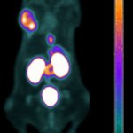
Hybrid PET/CT imaging can fully play to its strengths and steer treatment towards more effective procedures for diagnosing prostate cancer. The examination of the specific antigen PSMA with hybrid PET imaging enables treatment monitoring with significantly higher diagnostic accuracy than conventional imaging and therefore, Professor Clemens Cyran believes, will soon become the standard diagnostic…
Researchers at the University of Arizona are developing a Covid-19 testing method that uses a smartphone microscope to analyze saliva samples and deliver results in about 10 minutes. The research team, led by biomedical engineering professor Jeong-Yeol Yoon, aims to combine the speed of existing nasal swab antigen tests with the high accuracy of nasal swab PCR, or polymerase chain reaction,…
Olympus Corporation announced that it has entered into an agreement to acquire Quest Photonic Devices B.V. for up to EUR50 million including milestone payments to strengthen its surgical endoscopy capabilities. Quest offers advanced fluorescence imaging systems (FIS) for the medical field, enabling more surgical endoscopy capabilities, compared to conventional imaging technologies.
Blood draws are no fun. They hurt. Veins can burst, or even roll — like they’re trying to avoid the needle, too. Oftentimes, doctors use blood samples to check for biomarkers of disease: antibodies that signal a viral or bacterial infection, such as SARS-CoV-2, the virus responsible for COVID-19, or cytokines indicative of inflammation seen in conditions such as rheumatoid arthritis and…
Researchers at the Francis Crick Institute and the University of Western Australia have developed a new imaging method to see where antibiotics have reached bacteria within tissues. The method could be used to help develop more effective antibiotic treatments, reducing the risk of antibiotic resistance.
Scientists at UC Berkeley and Gladstone Institutes have developed a new CRISPR-based COVID-19 diagnostic test that, with the help of a smartphone camera, can provide a positive or negative result in 15 to 30 minutes.
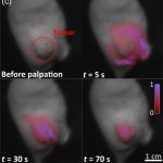
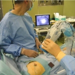
Italian neurosurgeon Professor Francesco Di Meco, explored the current and potential role of intra-operative ultrasound in neurosurgical oncology during the annual meeting of the European Association of Neurosurgical Societies (EANS) this October. The extent of resection is considered a prognostic factor in operative neuro oncology surgery and image-guided surgery is being regarded as one of the…
Researchers at Heriot-Watt University have developed a new technique that will allow medical professionals to see disease deep inside the human body in 10 times more detail. Professor Robert Thomson and his team want to improve endoscopies, when long, thin optical fibres are used to look inside the body. The new technique will open up a route to unprecedented imaging resolution, says Thomson, and…
Recent research in Clustered Regularly Interspaced Short Palindromic Repeats (CRISPR) has identified two enzymes that can detect Covid-19 RNA as simply as a pregnancy test Jesús Pla, an eminent microbiologist at the Complutense University in Madrid, explained in our exclusive interview. CRISPR technology could help alleviate workloads in packed hospitals and expand testing to primary care and…
‘Mini-lungs’ grown from tissue donated to Cambridge hospitals has provided a team of scientists from South Korea and the UK with important insights into how COVID-19 damages the lungs. Writing in the journal Cell Stem Cell, the researchers detail the mechanisms underlying SARS-CoV-2 infection and the early innate immune response in the lungs.

Researchers at the University of Helsinki and the Dana-Farber Cancer Institute have identified the mechanism behind bone marrow failure developing in children that suffer from Fanconi anaemia. The findings will help to develop new therapies for the disorder.
As COVID-19 continues to spread, bottlenecks in supplies and laboratory personnel have led to long waiting times for results in some areas. In a new study, University of Illinois, Urbana-Champaign researchers have demonstrated a prototype of a rapid COVID-19 molecular test and a simple-to-use, portable instrument for reading the results with a smartphone in 30 minutes, which could enable…
Researchers studying tissue removed from patients noses during surgery believe they may have discovered the reason why so many people with COVID-19 lose their sense of smell, even when they have no other symptoms. In their experiments they found extremely high levels of angiotensin converting enzyme II (ACE-2) only in the area of the nose responsible for smelling. This Enzyme is thought to be the…
Using highly complex analytical techniques, a group of researchers from Charité – Universitätsmedizin Berlin were able to observe in detail how different metals are released from joint implants and accumulate in the surrounding bone tissue. Findings showed a steady release of metals from various implant components. In contrast to previous assumptions, this was not related to the degree of…
Scientists from Nanyang Technological University, Singapore (NTU Singapore) and the Agency for Science, Technology and Research (A*STAR) have showed that applying “temporal pressure” to the skin of mice can create a new way to deliver drugs. In a paper published in Science Advances, the researchers showed that bringing together two magnets so that they pinch and apply pressure to a fold of…
Researchers have developed a novel three-dimensional imaging system to diagnose cervical cancer faster, non-invasively and more efficiently than conventional methods.
Researchers at Hokkaido University have succeeded in detecting anti-avian influenza virus antibody in blood serum within 20 minutes, using a portable analyzer they have developed to conduct rapid on-site bio tests. If a suitable reagent is developed, this technology could be used to detect antibodies against SARS-CoV-2, the causative virus of COVID-19.
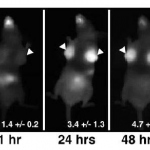
New imaging technique combines two technologies to spot early warning signs of Alzehimer's disease in mice.

Despite the use of personal protective equipment (PPE), reports show that many health care workers contracted the coronavirus disease (COVID-19), which raises substantial concerns about the effectiveness of the PPE.
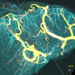
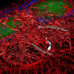
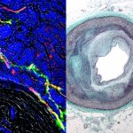
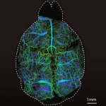
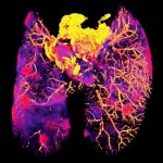
Diseases of the brain are often associated with typical vascular changes. Now, scientists at LMU University Hospital Munich, Helmholtz Research Centre for Environmental Health and the Technical University of Munich (TUM) have come up with a technique for visualising the structures of all the brain's blood vessels – right down to the finest capillaries – including any pathological changes. So…
Glioblastomas are complex, fast-growing malignant brain tumors that are made up of various types of cells. Even with aggressive treatment — which often includes surgery, radiation, and chemotherapy — glioblastomas are difficult to treat, leading to an average survival of 11-15 months. In research published in Science Advances, Xavier Intes, a professor of biomedical engineering at Rensselaer,…
Teams at Helmholtz Zentrum München, LMU Munich and the Technical University of Munich (TUM) have developed a new algorithm that enables automated detection of metastases at the level of single disseminated cancer cells in whole mice. Cancer is one of the leading causes of death worldwide. More than 90% of cancer patients die of distal metastases rather than as a direct result of the primary…
Johns Hopkins engineers have created a new lens-free ultra-miniaturized endoscope, the size of a few human hairs in width, that is less bulky and can produce higher quality images. Their findings were published in Science Advances. “Usually, you have sacrifice either size or image quality. We’ve been able to achieve both with our microendoscope,” says Mark Foster, an associate professor of…
Inhalation therapy is widely used for the treatment of lung diseases. Targeting of drugs to the site of disease is a major goal to improve drug efficacy and minimize side effects. Scientists at the Helmholtz Zentrum München and the Technical University of Munich have now shown that combined insight from various imaging methods allows for real-time monitoring of the dynamic process of drug…
Scientists at St George’s, University of London, in collaboration with researchers from Germany, the USA, Tunisia and Iran have identified a new gene associated with the neuromuscular disorder, hereditary spastic paraplegia (HSP). The study, published in Nature Communications, also highlights a potential mechanism for the disease, which is already being targeted in drug trials for Alzheimer’s…
The potential impact of undiagnosed sexually transmitted chlamydia infection on men’s fertility has been highlighted in an Australian-led study, which for the first time found chlamydia in the testicular tissue biopsies of infertile men whose infertility had no identified cause. The researchers from Queensland University of Technology also found antibodies specific to the bacteria responsible,…
Viscosity sensors are used by scientists fighting various diseases, such as Alzheimer's and Diabetes. Vilnius-based chemist Aurimas Vyšniauskas has enhanced their measuring capabilities. This breakthrough will equip researchers with additional tools to study living cells and how they change in a sick organism.
Cells are the basic building blocks of life – and, as such, they have been the object of intense study since the invention of the optical microscope in the 17th century. The development of mass spectrometry (MS) methods – those which define the chemical composition of cells – represented a further milestone for research in the field of cell biology. In the latest issue of the journal Nature…
Scientists from EPFL have developed an algorithm that can determine whether a super-resolution microscope is operating at maximum resolution based on a single image. The method is compatible with all types of microscopes and could one day be a standard feature of automated models.
A little bit of norovirus – the highly infectious microbe that causes about 20 million cases of food poisoning in the United States each year – goes a long way. Just 10 particles of the virus can cause illness in humans. A team of University of Arizona researchers has created a simple, portable and inexpensive method for detecting extremely low levels of norovirus. Jeong-Yeol Yoon, a…

In terms of success in revolutionary cancer treatment, molecular genetic examination procedures have developed immensely over recent years. They now range from conventional polymerase chain reactions (PCR) or fluorescence-in-situ hybridisation (FISH) to Next Generation Sequencing (NGS) with analysis of the entire exome or genome (Whole-Exome, WES or Whole-Genome, WGS) and of the transcriptome…
Kiel biochemistry research team proves the existence of a previously unknown alternative cholesterol transport mechanism inside cells. Cholesterol is a vital cell building block in humans and animals, and an integral part of the so-called cell membrane. This boundary layer separates the interior of the cell from the neighbouring cells and the surrounding environment. By means of certain proteins,…
Olympus has broken down barriers to imaging quality with the launch of its next-generation objectives. The breakthrough polishing technique enables the company to produce ultra-thin lenses that overcome the traditional trade-off between numerical aperture (NA), flatness and chromatic correction – enabling all three parameters to be significantly improved. Olympus has harnessed this proprietary…

Oxford Gene Technology (OGT), A Sysmex Group Company, has celebrated the opening of its new facility in Cambridge, UK. The opening ceremony, which took place at the company’s new premises on the prestigious Cambridge Science Park, was attended by the Department for International Trade (DIT) and local media as well as top-level representatives from OGT and its parent company, Sysmex Corporation.…

The critical element of testing for sepsis lies not so much in the location but in the timing and rapidity of results, according to Professor Jeannine T. Holden from Beckman Coulter Early identification enables treatment protocols to be delivered more quickly, offering better patient outcomes. Those most at risk, suggests Holden, are not patients within the intensive care unit – who are already…

Using urine to obtain diagnostic insights has been done for thousands of years and still remains an important tool to obtain crucial information. Covering a range of tests, urinalysis may be used to screen for or help to diagnose ailments such as urinary tract infections, kidney disorders, liver problems, diabetes or other medical conditions, just to name a few. Because urinalysis has been around…

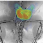
A novel imaging technique that uses a synthesized form of scorpion venom to light up brain tumors has shown promise in a clinical trial. The imaging system enables neurosurgeons to better see malignant growths that often are difficult to fully eliminate.
Body painting is considered by some to be the most ancient form of art. Its origins stem from tribal cultures, where its use was for ritual and ceremony. Today it is a familiar sight at carnivals and sporting events. It is also a frequently observed activity within the anatomy classrooms of medical schools around the world. But now lecturers at Hull York Medical School are pioneering the use of…
Until now, researchers haven’t been able to accurately quantify a latent form of HIV that persists in patients’ immune cells. A new genetic technique is fast and 10 to 100 times more accurate than previous diagnostics. This hidden, inactive version of HIV embeds into cells’ genomes and can persist despite otherwise successful therapies – thwarting attempts to cure the infection. Using a…
A team of University of Illinois at Urbana-Champaign researchers led by Bioengineering Professor Stephen Boppart has successfully visualized the tumor microenvironment of human breast tissue shortly after it was surgically removed from a patient in the operating room. The researchers achieved this using a new portable optical imaging system developed in Boppart's lab.

A research team led by Tufts University engineers has developed a non-invasive method for detecting bladder cancer that might make screening easier and more accurate than current invasive clinical tests involving visual inspection of bladder. In the first successful use of atomic force microscopy (AFM) for clinical diagnostic purposes, the researchers have been able to identify signature features…
This could be a major step towards a better understanding of the functions of deeply hidden brain compartments, such as the formation of memories, as well as related dysfunctions, including Alzheimer’s disease. Researchers from the Leibniz Institute of Photonic Technology (Leibniz IPHT) in Jena and the University of Edinburgh have succeeded in using a hair-thin fibre endoscope to gain insights…
Accelerate your workflow with the intelligent, high-resolution DP74 brightfield and fluorescence camera from Olympus. Histologists and pathologists face intense, rushed workloads where every minute counts. Experience enhanced efficiency with our next-generation camera. The DP74 creates a map of where you’ve been and can easily and rapidly lead you back to previous positions on the sample,…
Nanosystems that deliver anticancer drugs or imaging materials to tumours are showing significant progress, particularly those that respond to tumour-related stimuli, according to a review published in the journal Science and Technology of Advanced Materials. However, further research is still required to make sure these delivery systems are stable, non-toxic and biodegradable. Nanocarriers…
Lab-on-a-chip devices harness electrical signals to measure glucose, discern blood type and detect viruses or cancer. But biological samples need hafnium oxide for protection from electric fields.
While the role of the laboratory in disease diagnosis and management has expanded in recent years, causing an overwhelming rise in testing demands, the availability of skilled technologists and specialists has been diminishing. To meet the needs of an overworked and increasingly generalized workforce, today’s products not only must deliver more clinical data than ever before, but also must be…
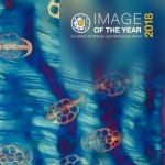
Olympus’ Image of the Year Award for light microscopy in Europe recognizes the very best in life science imaging. Inspired by the beauty and breadth of images submitted for Image of the Year 2017, Olympus is now continuing its quest for the best light microscopy art in 2018. For the chance to win one of three prizes, applicants can submit life science light microscopy images to…
Babraham Institute researchers have shown that some tumours use not one but two levels of protection against the immune system. Knocking out one level boosted the protective effects of the second and vice versa. The research demonstrates that a two-pronged approach targeting both cell types simultaneously may offer a promising route for the development of new cancer immunotherapies.
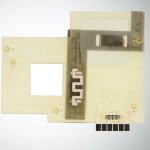
A thin copper wire wrapped around a channel slightly thicker than a strand of hair could be the key to manufacturing a compact electronic device capable of counting white blood cells from the comfort of one’s home, a Kennesaw State University researcher says. Hoseon Lee, an assistant professor of electrical engineering in the Southern Polytechnic College of Engineering and Engineering…
Recognising malignant tissue remains a tricky task. While today, most patients undergo a biopsy, an invasive procedure where tissue is sampled, stained and assessed, researchers are exploring the potential of optical biopsy, the visual assessment of suspect tissue. The interest in optical biopsy ‘is indeed enormous,’ confirms Dr Thomas Bocklitz, physicist at Friedrich-Schiller University in…
Indiana University researchers have made an important step forward in the design of drugs that fight the hepatitis B virus, which can cause liver failure and liver cancer. It's estimated that 2 billion people worldwide have had a hepatitis B virus infection in their lifetime, with about 250 million -- including 2 million Americans -- living with chronic infection. Although a vaccine exists, there…

Machine learning is playing an increasing role in computer-aided diagnosis, and Big Data is beginning to penetrate oncological imaging. However, some time may pass before it truly impacts on clinical practice, according to leading UK-based German researcher Professor Julia Schnabel, who spoke during the last ESMRMB annual meeting. Machine learning techniques are starting to reach levels of human…
A powerful antiviral protein may act as a checkpoint for keeping or ending a pregnancy. When exposed to Zika virus before birth, mouse fetuses with the protein commit cell suicide, while fetuses without it continued to develop. The result, published in Science Immunology, suggests that the protein, a receptor involved in immune cell signaling, plays a role in spontaneous abortions and other human…
Smith & Nephew, the global medical technology business, announces the European launch of MolecuLight i:X, the easy to use, handheld imaging device that instantly measures wound surface area and visualises the presence and distribution of potentially harmful bacteria in wounds. Currently wound assessments are made with the naked eye which can lack the accuracy required to most effectively…
Nanosensors are incredible information-gathering tools for myriad applications, including molecular targets such as the brain. Neurotransmitter molecules govern brain function through chemistry found deep within the brain, so University of California, Berkeley researchers are developing nanosensors to gain a better understanding of exactly how this all plays out. During the AVS 64th…
Columbia University Medical Center (CUMC) researchers have identified cells in the upper digestive tract that can give rise to Barrett’s esophagus, a precursor to esophageal cancer. The discovery of this “cell of origin” promises to accelerate the development of more precise screening tools and therapies for Barrett’s esophagus and esophageal adenocarcinoma, the fastest growing form of…

For many years, we thought that all proteins must fold into complicated shapes to fulfill their functions, looking like thousands of sets of custom-tailored locks and keys. But over the past two decades, scientists have begun to realize other proteins—including those involved in many essential cellular functions—remain fully or partially unfolded for parts of their lives. Out of this…
Surgeons at Penn Medicine are using a fluorescent dye that makes cancerous cells glow in hopes of identifying suspicious lymph nodes during head and neck cancer procedures. Because the tumors glow, surgeons get real-time guidance to help them take out as much cancer as possible and leave non-diseased tissue alone.
In a sharp and pointy world, wound healing is a critical and marvelous process. Despite a tremendous amount of scientific study, many outstanding mysteries still surround the way in which cells in living tissue respond to and repair physical damage. One prominent mystery is exactly how wound-healing is triggered: A better understanding of this process is essential for developing new and improved…
Certain members of Generation Y, who grew up alongside enormous information technology (IT) advances, now occupy decision-making roles. Meanwhile, generation Z is emerging into the continuing IT revolution; the digital world surrounds us.
Columbia University Medical Center researchers have found that it may be possible to access memories “lost” to Alzheimer’s disease, if their discoveries about memory loss in mice also apply to people with the disease.

Scientists from the University of Würzburg have synthesized a complex sugar molecule which specifically binds to the tumor protein Galectin-1. This could help to recognize tumors at an early stage and to combat them in a targeted manner.
The reasons why doctors request urinary analysis are varied – perhaps to detect a possible or suspected infection, or to screen for kidney diseases. In all cases a reliable and rapid result is the major aim. Urinary microscopy and culture have been the mainstays of urinary analysis for many, many years both of which require time and specialist handling.

Genes do not exist in isolation. So far, little has been known about how the position of a gene on a chromosome affects its evolution.
A saliva test developed by researchers at the Johns Hopkins Bloomberg School of Public Health nearly matches the performance of a blood test widely used to assess recent or past hepatitis E virus infections, a new study reports. The findings could offer an easier, less expensive alternative to gathering data for studying and eventually treating the disease, which infects an estimated 20 million…
Olympus’ new BX53 microscope provides bright, sharp images with excellent color rendering performance equivalent to halogen lamps. The long-life LED light source, the True Color LED, is brighter and more uniform than a 100-watt halogen bulb – matching every contrast method and providing bright images to multi-head discussion systems for up to 26 people.
Canadian researchers have invented an intraoperative probe that reliably detects multiple types of tumour cells.
Today, tracking the development of individual cells and spotting the associated factors under the microscope is nothing unusual. However, impairments like shadows or changes in the background complicate the interpretation of data. Now, researchers at the Technical University of Munich (TUM) and the Helmholtz Zentrum München have developed a software that corrects images to make hitherto hidden…
Researchers at the Institute of Bioengineering and Nanotechnology (IBN) of A*STAR have engineered a three-dimensional heart tissue from human stem cells to test the safety and efficacy of new drugs on the heart.

Oxford Gene Technology (OGT), The Molecular Genetics Company, today announced that it has signed an agreement to be acquired by Sysmex Corporation, a Japanese in vitro diagnostic company.
Light microscopy today offers a wealth of techniques that provide fascinating insights into life on subcellular level. “In light microscopy these days there are so many new techniques that each of us can only handle a subset of them,” says Christian Tischer, scientific officer in der Advanced Light Microscopy Facility of the European Molecular Biology Laboratory (EMBL) in Heidelberg, Germany,…
Study reveals the surprising role of omega-3 fatty acids in keeping the blood-brain barrier closed
Ideal for high-throughput, regular use, Olympus’ ergonomic CX43 and CX33 microscopes deliver superior comfort and reduce fatigue during long periods of routine microscopy.

A simple eye test could help solve the biggest global cause of irreversible blindness, glaucoma. In clinical trials, the pioneering diagnostic test - developed by researchers at UCL Institute of Ophthalmology and the Western Eye Hospital - allowed doctors to see individual nerve cell death in the back of the eye.
Autonomous driving, automatic speech recognition, and the game Go: Deep Learning is generating more and more public awareness. Scientists at the Helmholtz Zentrum München and their partners at ETH Zurich and the Technical University of Munich (TUM) have now used it to determine the development of hematopoietic stem cells in advance. In ‘Nature Methods’ they describe how their software…
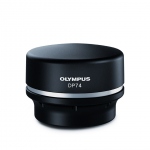
Helping to save time and increase comfort at the microscope, Olympus’ new DP74 camera delivers intelligent imaging with a range of innovative features designed to facilitate the workflow and enhance return on investment (ROI).
An international group of researchers report success in mice of a method of using positron emission tomography (PET) scans to track, in real time, an antibody targeting a hormone receptor pathway specifically involved in prostate cancer.
Bladder cancer (BC) is the most common malignancy involving the urinary system and the ninth most common malignancy worldwide.

The increasing numbers of bacteria resistant to the newer generations of antibiotics is a public health problem on a global scale. Bacteria have an extraordinary capacity for adaptation, mutating permanently to overcome the action of our increasingly impotent antimicrobial armamentarium. A situation further aggravated by the use of the powerful ‘large spectrum’ antibiotics, creating further…
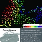
Nanoparticles known as Cornell dots, or C dots, have shown great promise as a therapeutic tool in the detection and treatment of cancer. Now, the ultrasmall particles – developed more than a dozen years ago by Ulrich Wiesner, the Spencer T. Olin Professor of Engineering at Cornell University – have shown they can do something even better: kill cancer cells without attaching a cytotoxic drug.
NIBIB researchers have created a nanovaccine that could make a current approach to cancer immunotherapy more effective while also reducing side effects. The nanovaccine helps to efficiently deliver a unique DNA sequence to immune cells – a sequence derived from bacterial DNA and used to trigger an immune reaction. The nanovaccine also protects the DNA from being destroyed inside the body, where…
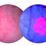
Researchers at the Cancer Research UK Cambridge Institute sprayed a dye on oesophageal tissue samples taken from people with Barrett’s oesophagus – a condition that increases the risk of developing oesophageal cancer. The dye sticks to healthy oesophageal cells but not to pre-cancerous cells. They then shone near-infrared light - which is just beyond the red colours that our eyes can normally…

Cold plasma looks like the glow from the “Star Wars” blue light saber but this beam of energy, made of electrons that change polarity at micro-second or nanosecond speeds, could help bones heal faster, according to researcher from the Thomas Jefferson University.
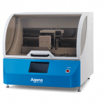
Cancer patients today can benefit from much better drugs providing treatments tailor-made to specific mutations. However, broad application is still hampered by a considerable bottleneck: fast, reliable, and cost-effective diagnosis. Agena's MassARRAY platform is addressing this bottleneck with an elegant new solution.
As part of a national, joint research project in cooperation with Chronix Biomedical (San Jose, CA/USA/Göttingen/Germany), Professor Michael Oellerich MD is on new biomarkers in organ transplantation, aiming to develop personalised immunosuppression for patients. This also entails the development of molecular test procedures, among others for the early detection of rejection. The keyword here is…
UK researchers have identified a new approach, which could make it easier to identify fatty plaques that could cause heart attacks or a stroke.
When a woman is diagnosed with the earliest stage of breast cancer, how aggressive should her treatment be? Will the non-invasive cancer become invasive? Or is it a slow-growing variety that will likely never be harmful? Researchers at the University of Michigan developed a new technology that can identify aggressive forms of ductal carcinoma in situ, or stage 0 breast cancer, from non-aggressive…
Two new optical devices could reduce the need to take tissue samples during medical examinations and operations and to then send them for testing – potentially speeding up diagnosis and treatment and cutting healthcare costs.

Histopathologists play key roles in diagnosing disease entities and determining biomarkers related to the prognosis and response to specific therapy of malignant tumours. Report: Bela Molnar

Strictly speaking, digital pathology has not yet resulted in any groundbreaking changes for clinical diagnostics. The conventional light microscope introduced to pathology around 100 years ago continues to be the most important tool for pathologists.
Although tuberculosis (TB) is commonly thought of as being a disease that mainly affects nineteenth century poets and Victor Hugo characters, it is still the second-most common cause of mortality from an infectious disease in the world, killing nearly three people every minute.
The ongoing debate about breast cancer diagnostics has left many women confused — particularly over what age they should get mammograms and who needs treatment. An issue with current methods is that they often identify lumps but cannot conclusively pinpoint which ones are cancerous. To help resolve this uncertainty, researchers have developed a pill that could improve imaging techniques so that…
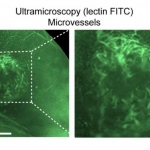
Stopping the growth of blood vessels in tumours is a key target for glioblastoma therapies, and imaging methods are essential for initial diagnosis and monitoring the effects of treatments. While mapping vessels in tumours has proven a challenge, researchers have now developed a combined magnetic resonance imaging (MRI) and ultramicroscopy 'toolkit' to study vessel growth in glioma models in more…
Scientists of the Göttingen Cluster of Excellence CNMPB and the EMBL describe efficient fluorescence tag for super-resolution microscopy.
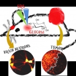
Researchers have identified a mechanism that allows cancer cells to respond and grow rapidly when levels of sugar in the blood rise. This may help to explain why people who develop conditions in which they have chronically high sugar levels in their blood, such as obesity, also have an increased risk of developing certain types of cancer.
Doctors at the Duke University School of Medicine have tested a new injectable agent that causes cancer cells in a tumor to fluoresce, potentially increasing a surgeon’s ability to locate and remove all of a cancerous tumor on the first attempt. The imaging technology was developed through collaboration with scientists at Duke, the Massachusetts Institute of Technology (MIT) and Lumicell Inc.
Proteins are like a body’s in-house Lego set. These large, complex molecules are made up of building blocks called amino acids. Most of the time, proteins fold correctly, but sometimes they can misfold. This misfolding causes the proteins to get sticky, and that can promote clumping, or aggregation, which is the hallmark of several neurodegenerative diseases such as ALS, Alzheimer’s and…
Antibodies combat viruses and bacteria. They also attach themselves to cancer cells – in a typical, characteristic way. Fraunhofer scientists are using this property to detect cancer cells in tissue samples. Such rapid tests can already be applied by surgeons during operations – within a few minutes and without expensive equipment.

Scientists have shown for the first time that an enzyme crucial to keeping our immune system healthy “surfs” along the strands of DNA inside our cells. The researchers used extremely powerful microscopy to watch how the enzyme AID (activation-induced deoxycytidine deaminase) moves around and interacts with other molecules.

Bladder cancer is associated with high recurrence rates, necessitating prolonged surveillance and repeated treatments. As a result, it is one of the most challenging and costly of all solid tumours to manage. Although most patients present at an early stage with non-muscle-invasive bladder cancer (NMIBC), between 13% and 61% will experience recurrence within 1 year of initial transurethral…
One goal of immunotherapy is to rally a patient’s often over-burdened immune cells to effectively attack a tumor. Among foot soldiers on the immune front line is a subpopulation of white blood cells called “patrolling monocytes,” whose job is to cruise the bloodstream, cart off cellular debris, and block invasion of a less benign population of inflammatory cells.
When it comes to fending off disease and helping prevent people from falling ill, the body’s immune system – armed with T-cells that help eliminate cancer cells, virus-infected cells and more – is second to none. But exactly how the immune system works remains, in many ways, a mystery, as there are numerous cell types whose functions and interactions with our immune systems have not been…
A team of scientists at the Children’s Research Institute at UT Southwestern (CRI) has become the first to use a tissue-clearing technique to localize a rare stem cell population, in the process cracking open a black box containing detailed information about where blood-forming stem cells are located and how they are maintained. The findings provide a significant advance toward understanding…
Researchers at the Virginia Bioinformatics Institute at Virginia Tech have uncovered key cellular functions that help regulate inflammation -- a discovery that could have important implications for the treatment of allergies, heart disease, and certain forms of cancer.
Automated image analysis shows significant potential within histopathology to help identify novel and subtle prognostic features. UK expert Dr Peter Caie also believes such image analysis can turn aspects of histopathology from a traditionally semi-quantitative field into a fully quantifiable and standardised science. However, he also points out that challenges remain before the full potential is…
Stony Brook researchers publish experimental findings in the Journal of Neuroscience that show the lateral position more efficiently rids the brain of solutes that may contribute to disease.
Founded in 1968, The Sysmex Corporation has challenged its way past far larger companies to become one of the leading healthcare companies around the world. How? By focusing constantly on one objective: maximising the strength of our people, knowledge and resourcefulness to live up to our mission statement: Shaping the Advancement of Healthcare.
Researchers at Columbia University have reported a new approach to visualize glucose uptake activity in single living cells by light microscopy with minimum disturbance.

Seegene Inc. today announced that it has entered into a worldwide collaboration agreement with QIAGEN N.V. to develop and supply molecular diagnostic assays.

The view across the Atlantic – it fills Professor Fabian Kiessling, Chair of Experimental Molecular Imaging at the RWTH Aachen (Rhine-Westphalia Institute of Technology Aachen), with optimism. The USA offers more opportunities for molecular imaging. Only recently, new tracers for Alzheimer’s were accepted as reimbursable in some centres, whilst the development of new diagnostics in Europe…

New research in The FASEB Journal suggests that clinical trials of omega-3 and antioxidant supplementation should be undertaken for people with Alzheimer's disease with mild clinical impairment.
Researchers at the University of Toronto design diagnostic chip to reduce testing time from days to one hour, allowing doctors to pick the right antibiotic the first time.
Genetically engineered fibers of the protein spidroin, which is the construction material for spider webs, has proven to be a perfect substrate for cultivating heart tissue cells, MIPT researchers found. They discuss their findings in an article that has recently come out in the journal PLOS ONE.
Gideon Ho, CEO and co-founder of Singapore-based HistoIndex is confident: ‘After a biopsy a patient waits in a hospital bed, but now, instead of waiting a couple days until doctors know how to treat this patient, we can deliver results while the patient is still in the hospital.’ Report: John Brosky
Trichomonias, with an estimated 187 million cases, and Chlamydia with around 100 million, are the most prevalent sexually transmitted diseases (STDs), according to the World Health Organisation (WHO). There are approximately 36 million cases each of gonorrhoea and syphilis. HIV1/2 cases are around 34 million. Report: Cynthia Keen
A tiny nanoscale device can accurately measure a patient’s blood for methotrexate – a commonly used but potentially toxic cancer drug – in under 60 seconds, according to biomedical instrument designer Jean-François Masson, and Joelle Pelletier, a DHFR enzyme specialist, both at the Chemistry Department, University of Montreal.

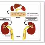
Donor transplant rejection and cancer relapse have two things in common: early recognition is vital and monitoring is hugely challenged.
Researchers from the University of Heidelberg and the German Cancer Research Center (DKFZ) have developed a new method that uses light to control processes in living cells.
A recent study from the Heidelberg-based company Sciomics, a spin-off from scientists from the German Cancer Research Center (DKFZ), has presented an advanced method to predict the recurrence of bladder cancer after surgery. The method, which can help avoid frequent cystoscopy examinations in a majority of patients, is based on an analysis of the protein composition of cancer tissue obtained…
Olympus launches its OlyVIA Mobile iPad App, an image viewer dedicated to virtual slides.

Medical technologies have maintained a leading position in European patent registrations for 15 years.

Two statements from publications by Dr Stephanie Dancer, Department of Microbiology, Hairmyres Hospital, East Kilbride (UK) prompted Ralf Mateblowski to interview Professor Markus Dettenkofer, Acting Director of the Institute for Environmental Medicine and Hospital Hygiene, Freiburg University Medical Centre about environmental and infection control
A British teenager, 17-year-old Fred Turner, has won a major accolade for building a DNA testing analyser in his own home for a few hundred euros.

Apart from tumour immunologists themselves, for a long time oncologists have underestimated the role of the immune system in cancer treatment. Nonetheless, in recent years increasing attention has been given to this aspect of cancer.

Olympus VS120 virtual slide scanning system earns three awards at the prestigious second International Scanner Contest.
Photoacoustics offers an additional contrast mechanism that makes the technology far more sensitive for the presence of blood.

Catastrophes draw people closer, as demonstrated by the development of the new high-end ultrasound scanner Aplio 500 from Toshiba. The clinical evaluation period took place during the tsunami and the nuclear catastrophe in Fukusima. Professor Thomas Fischer at the Radiological Institute, Charité Clinic in Berlin, was impressed by the enormous commitment shown by the Japanese firm’s engineers…
Breast cancer is the most common form of cancer in women. Evaluation of the axillary lymph nodes is essential to insure complete cancer removal. Fluorescence imaging instead of radioisotopes is an innovative method for sentinel lymph node detection.
Scientists at the University of Strathclyde in Glasgow are developing a technique based on a new discovery which could pave the way towards detecting Alzheimer’s disease in its earliest stages - and could help to develop urgently-needed treatments.

A new technique to aid the early detection and diagnosis of inflammatory bowel disease has been developed by UK and Germans researchers. The Confocal Laser Endomicroscope (CLE) contains a powerful microscope that allows clinicians to view bacteria in the intestine that are thought to trigger bowel diseases such as Crohn’s and ulcerative colitis.
It sounds like an idea plucked from the realms of science fiction writing. But in this case, there is nothing fictional about it. Scientists in Yorkshire have developed a process that uses the luminous cells from jellyfish to diagnose cancers deep within the human body.

7,000 people from 120 countries met in Stockholm this September to hear international experts discuss the progress, solutions and challenges of one of our greatest healthcare burdens. Prevention, self-monitoring, surgery, guidelines, economic problems, drug-safety, and co-morbidities – these are just a few of the problems associated with the care of about 55 million diabetics in Europe.
Molecular imaging, the discipline that unites molecular biology and in vivo imaging technologies to assess biological activity in the body, promises to open up ‘…an entire new universe,’ declared Dr Ralph Weissleder, of the Centre for Molecular Imaging Research at Massachusetts General Hospital, USA, in the journal Radiology. That was just one decade ago. And he was right. It has indeed…
Molecular imaging, the discipline that unites molecular biology and in vivo imaging technologies to assess biological activity in the body, promises to open up ‘…an entire new universe,’ declared Dr Ralph Weissleder, of the Centre for Molecular Imaging Research at Massachusetts General Hospital, USA, in the journal Radiology. That was just one decade ago. And he was right. It has indeed…

Molecular imaging, a new science that emerged from molecular biology, is unlike traditional imaging. Whilst the latter can, for example, show the differences in proton density or water content on MRI, molecular imaging uses biomarkers (probes) that interact selectively with molecules within an area and then generate the image according to fine molecular alterations occurring inside (e.g. within a…
The detection and documentation of low light fluorescence signals in live cell experiments is a particular challenge for digital cameras. To meet this demand, Leica Microsystems adds the new Leica DFC345 FX to its portfolio of powerful digital cameras. In addition to combining high sensitivity with high resolution, this new camera features a fast image capture rate and a broad dynamic range,…
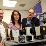
The Innovation Prize for outstanding, application-oriented ideas in life sciences has been awarded by the Working Group of BioRegions at the Biotechnica in Hanover to research groups from Heidelberg, Munich and Ulm. Professor Lisa Wiesmüller, of the Women’s Hospital, University of Ulm, received the €2,000 prize for developing a test system for the identification and early detection of breast…
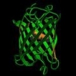
Professor Roger Tsien, Nobel Laureate in Chemistry 2008 for the ‘Discovery and Development of Green Fluorescent Proteins’, was one three* Keynote Speakers at the opening ceremony of the Medical Physics and Bioengineering World Congress 2009, held in Munich this September.

For the past 30 years, in the former USSR and Russia, laser medicine has been actively developed and regular annual conferences have opened new avenues for its use by the country's doctors.
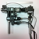
The new Leica LMD7000 is a laser microdissection system with a power-adjustable, high precision laser.
The Leica LMD7000 is the only laser microdissection system with a power adjustable, high precision laser. For the first time, high laser power and high repetition rates, are combined within one system. The laser's high pulse repetition rates are ideal for the fast excision of single cells, cell clusters, or thin and soft samples. Additionally, high laser power allows the dissection of thick or…