Tiny nanodevice monitors cancer treatment
A tiny nanoscale device can accurately measure a patient’s blood for methotrexate – a commonly used but potentially toxic cancer drug – in under 60 seconds, according to biomedical instrument designer Jean-François Masson, and Joelle Pelletier, a DHFR enzyme specialist, both at the Chemistry Department, University of Montreal.
Crucially, the device’s optical system can rapidly gauge the optimal dose of methotrexate a patient needs, while minimising the drug’s adverse effects.
Methotrexate can block the enzyme dihydrofolate reductase (DHFR), which is active in the synthesis of DNA precursors and thus promotes cancer cell proliferation – but it is highly toxic, so closely monitoring the drug’s concentration in a patient’s serum is vital.
Currently, a device using fluorescent bioassays to measure light polarisation produced by a drug sample is used – but this is a cumbersome, expensive platform that only experienced personnel can manipulate.
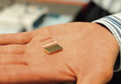
Six years ago, Pelletier and Masson investigated how to simplify the procedure. In the course of their research they developed and manufactured the miniaturised device that works by surface plasmon resonance. Put simply, it measures serum methotrexate concentration through gold nanoparticles on the surface of a receptacle. In ‘competing’ with methotrexate to block the enzyme, the gold nanoparticles change the colour of light detected by the instrument and that colour reflects the exact concentration of the drug in the blood sample.
In tests, measurements from nanoparticles device proved as accurate, Masson pointed out, yet took under 60 seconds to produce, compared to 30 minutes for current devices used, and they were obtained easily by lab technicians inexperienced in surface plasmon resonance.
The new device is small and needs little manipulation of samples, so could soon be used at the bedside and/or in a GP surgery.
10.11.2014