Credit: Mark Foster
News • Imaging the brain
Ultra-miniaturized endoscope produces HQ images
Johns Hopkins engineers have created a new lens-free ultra-miniaturized endoscope, the size of a few human hairs in width, that is less bulky and can produce higher quality images.
Their findings were published in Science Advances.
“Usually, you have sacrifice either size or image quality. We’ve been able to achieve both with our microendoscope,” says Mark Foster, an associate professor of electrical and computer engineering at The Johns Hopkins University and the study’s corresponding author. Intended for examining neurons firing off in the brains of animals such as mice and rats, an ideal microendoscope should be small to minimize brain tissue damage yet powerful enough to produce a clear image.
Currently, standard microendoscopes are about half a millimeter to a few millimeters in diameter, and require larger, more invasive lenses for better imaging. While lensless microendoscopes exist, the optical fiber within that scans an area pixel by pixel frequently bends and loses imaging ability when moved.
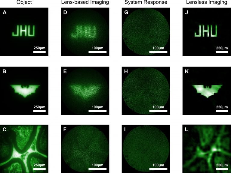
In their new study, Foster and colleagues created a lens-free ultra-miniaturized microendoscope that, compared to a conventional lens-based microendoscope, increases the amount researchers can see and improves image quality. The researchers achieved this by using a coded aperture, or a flat grid that randomly blocks light creating a projection in a known pattern akin to randomly poking a piece of aluminum foil and letting light through all of the small holes. This creates a messy image, but one that provides a bounty of information about where the light originates, and that information can be computationally reconstructed into a clearer image. In their experiments, Foster’s team looked at beads in different patterns on a slide.
Thanks to computational reconstruction, we can purposefully capture something that looks awful and counterintuitively end up with a clearer final image
Mark Foster
“For thousands of years, the goal has been to make an image as clear as possible. Now, thanks to computational reconstruction, we can purposefully capture something that looks awful and counterintuitively end up with a clearer final image,” says Foster.
Additionally, Foster and team’s microendoscope doesn’t require movement to focus on objects at different depths; they use computational refocusing to determine where the light originated from in 3 dimensions. This allows the endoscope to be much smaller than a traditional one that requires moving the endoscope around to focus.
Looking forward, the research team will test their microendoscope with fluorescent labeling procedures in which active brain neurons would be tagged and illuminated, to determine how accurately the endoscope can image neural activity.
Source: John Hopkins University
10.12.2019