Image source: Pétusseau et al., Journal of Biomedical Optics 2022 (CC BY 4.0)
News • New imaging method
Delayed fluorescence for effective surgical tumor removal
In the surgical treatment of cancer, distinguishing between tumors and healthy tissues is critical. Fluorescent markers can help to do this, by enhancing the contrast of tumors during surgery.
Some markers show a phenomenon called “delayed fluorescence” (DF) which relies on detecting “hypoxia” (or low oxygen concentration), a condition often presented by tumors. The real-time imaging of hypoxia can provide a high contrast between tumors and healthy cells. This can allow surgeons to remove the tumor effectively. However, real-time hypoxia imaging for surgical guidance has not yet been achieved.
In a new study published in Journal of Biomedical Optics (JBO), researchers proposed an optical imaging system that enables real-time imaging of tissue oxygen concentration for tumors presenting chronic or transient hypoxia. The team achieved this by making use of an endogenous molecule called protoporphyrin IX (PpIX), which exhibits DF in the red to near-infrared region. “This is a truly unique reporter of the local oxygen partial pressure in tissues. PpIX is endogenously synthesized by mitochondria in most tissues, and the particular property of DF emission is directly related to low microenvironmental oxygen concentration,” explains Brian Pogue, Chair of Medical Physics at University of Wisconsin-Madison, Adjunct Professor of Engineering Sciences at Dartmouth College, and senior author of the study. “Healthy cells will show little to no DF, because it is quenched in the presence of molecular oxygen.”
The results reported by Petusseau’s team suggest hypoxia imaging as an efficient approach to identifying tumors in cancer treatment
Frédéric Leblond
The technical challenge in detecting DF is due to its low intensity; background noise makes it difficult to detect without a single photon detector. The team overcame this problem using a highly sensitive time-gated imaging system, which allows signal detection within a specified time window only. This greatly reduces the background noise and enables a wide-field direct mapping of oxygen partial pressure (pO2) changes with the acquired DF signal. The result is real-time metabolic information, a useful map for surgical guidance.
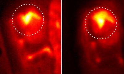
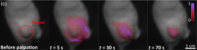
Lead author Arthur Petusseau, a doctoral candidate in Engineering Sciences at Dartmouth College, explains: “Acquiring both prompt and delayed fluorescence in a rapid sequential cycle allowed for imaging oxygen levels in a way that was independent of the PpIX concentration.” Petusseau’s team demonstrated the efficacy of their technique using mice models of pancreatic cancer, which exhibited hypoxic tumors. The DF signal obtained from the cancerous cells was over five times stronger than that from surrounding healthy oxygenated tissues. The signal contrast was further enhanced when the tissues were palpated before imaging to further enhance transient hypoxia.
According to Frédéric Leblond, Professor of Engineering Physics at Polytechnique Montréal and JBO Associate Editor, “The results reported by Petusseau’s team suggest hypoxia imaging as an efficient approach to identifying tumors in cancer treatment. PpIX DF detection uses a known clinical dye and an already-approved in-human marker, with great potential for surgical guidance, and more.” Petusseau notes that the imaging of pO2 in tissues could also enable control of tissue metabolism. This, in turn, would help us better understand the biochemistry involved in oxygen supply and consumption.
Source: SPIE – The International Society for Optics and Photonics
12.10.2022