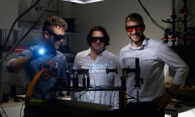
Source: Tomáš Čižmár
News • Photonic endoscopy
Fibre probe explores the depth of our brain
This could be a major step towards a better understanding of the functions of deeply hidden brain compartments, such as the formation of memories, as well as related dysfunctions, including Alzheimer’s disease. Researchers from the Leibniz Institute of Photonic Technology (Leibniz IPHT) in Jena and the University of Edinburgh have succeeded in using a hair-thin fibre endoscope to gain insights into hardly-accessible brain structures.
For the first time, scientists are able to achieve high-resolution observations of neuronal structures inside deep brain areas of living mice – by the use of the most minimally invasive endoscopic probe reported thus far. This study has been published in the current issue of the journal “Light: Science & Applications”.
For the first time, we have shown that it is possible to examine deep brain regions of a living animal model in a minimally invasive way and to achieve high-resolution images at the same time
Ivo T. Leite
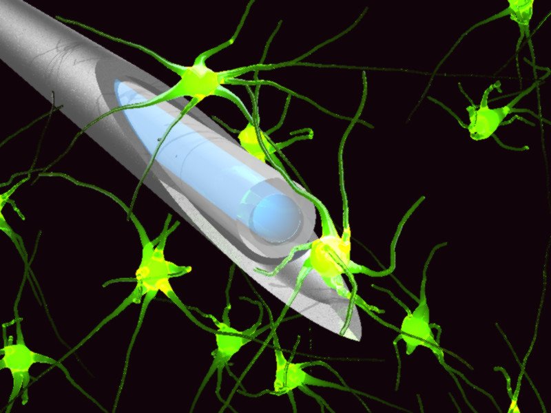
Using a hair-thin optical fibre, the researchers can look into deep brain areas of a living mouse as if through a keyhole. Recently introduced methods for holographic control of light propagation in complex media enable the use of a multimode fibre as an imaging tool. Based on this new approach, the scientists designed a compact system for fluorescence imaging at the tip of a fibre, offering a much smaller footprint as well as enhanced resolution compared to conventional endoscopes based on fibre bundles or graded-index lenses.
“We are very excited to see our technology making its first steps towards practical applications in neuroscience,” says Dr Sergey Turtaev from Leibniz IPHT, lead author of the paper. “For the first time, we have shown that it is possible to examine deep brain regions of a living animal model in a minimally invasive way and to achieve high-resolution images at the same time,” adds IPHT scientist Dr Ivo T. Leite. Sergey and Ivo work in the research group for Holographic Endoscopy led by IPHT scientist Prof. Tomáš Čižmár, who developed the holographic method for imaging through a single fibre. Using this approach, the research team succeeded in obtaining images of brain cells and neuronal processes in the visual cortex and hippocampus of living mice with resolution approaching one micrometre (i.e. one thousand times smaller than a millimetre). Detailed observations within these areas are crucial for research into sensory perception, memory formation, and severe neuronal diseases such as Alzheimer’s. Current investigation methods are strongly invasive, such that it is not possible to observe neuronal networks in these inner regions at work without massive destruction of the surrounding tissue – usual endoscopes comprised of hundreds of optical fibres are too large to penetrate such sensitive brain regions, while the neuronal structures are too tiny to be visualised by non-invasive imaging methods such as magnetic resonance imaging (MRI).
“This minimally invasive approach will enable neuroscientists to investigate functions of neurons in deep structures of the brain of behaving animals: without perturbing the neuronal circuits in action, it will be possible to reveal the activity of these neuronal circuits while the animal is exploring an environment or learning a new task,” explains project partner Dr Nathalie Rochefort from the University of Edinburgh.
Building up on this work, the research team now wants to address the current challenges of neuroscience, which will entail the delivery of advanced microscopy techniques through single fibre endoscopes. “Under the “Photonics for Life” flag of the Leibniz-IPHT and in the scope of the European Research Council funded project LIFEGATE, we will strive hard to prepare more significant advancements on this result, essentially funnelling the most advanced methods of modern microscopy deep inside the tissues of living and functioning organisms.” concludes Prof. Tomáš Čižmár.
Source: Leibniz Institute of Photonic Technology (Leibniz IPHT)
29.11.2018