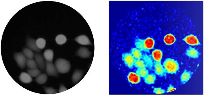
Image source: Heriot-Watt University
News • Improving imaging
Light up diseases with 'photonic lanterns'
Researchers at Heriot-Watt University have developed a new technique that will allow medical professionals to see disease deep inside the human body in 10 times more detail.
Professor Robert Thomson and his team want to improve endoscopies, when long, thin optical fibres are used to look inside the body. The new technique will open up a route to unprecedented imaging resolution, says Thomson, and the proof of concept is reported in the journal Nature Communications.
We’ve successfully demonstrated our technique can produce images an order of magnitude better than what’s currently possible, from microns to a few hundred nanometres
Robert Thomson
Thomson said: “Optical fibres are the ideal technology to look at diseases or other biological processes inside the body because they are thin and flexible. At the moment, the spatial resolution of the images they deliver is limited, because the optical fibres scramble the image if we try to push the resolution too high.”
Thomson and his team have now demonstrated that photonic lanterns will, in the future, dramatically improve what the fibre sees inside the body. “While photonic lanterns have been around for about a decade, nobody has ever applied them to an imaging application, or indeed a healthcare challenge like solving the limitations of micro endoscopies. We have shown that photonic lanterns allow us to manipulate light with exquisite precision, in a manner that is simply not possible with conventional approaches.”
When optical fibres transmit an image, the light is carried by rays of light, called modes, inside the fibres. In conventional optical fibres, the rays easily exchange light, a phenomenon called mode mixing: this scrambles the image. Thomson explained the difference: “The power of photonic lanterns is that they completely kill mode-mixing, and this means we can transmit exquisite images of structures from inside the body. We’ve successfully demonstrated our technique can produce images an order of magnitude better than what’s currently possible, from microns to a few hundred nanometres. With our technique, we hope to be able to see the shape of individual bacteria and other structures, instead of just blobs.”
Thomson and team are now working on securing funding so that they can demonstrate the full potential of the new approach and take the technology forward to commercialisation.
Source: Heriot-Watt University
25.11.2020