Boosting the potential of ultrasound
Photoacoustics offers an additional contrast mechanism that makes the technology far more sensitive for the presence of blood.
Absorption of pulsed light produces ultrasound signals. This photoacoustic approach to ultrasound imaging could help diagnose tumour growth and its suppression by therapy. An expert in Biomedical Photonic Imaging, Professor Wiendelt Steenbergen outlined the highly promising potentials of the technology during our European Hospital interview. Asked what the technical-medical principle is behind photoacoustic imaging, he explained: ‘Photoacoustic imaging is an ultrasound imaging modality where the ultrasound is generated by the absorption of short light pulses in the tissue. Absorption of pulsed light by e.g. haemoglobin and melanin causes a slight but rapid heating of the absorbing structures, leading to the emission of ultrasound. These waves are measured at the tissue surface and allow for constructing the 3-D distribution of optical absorption inside the tissue. The images therefore reveal blood vessels, enhanced blood concentration, but also optical contrast agents.’
How does this expand the horizons of imaging?
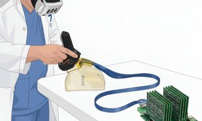
[Bild-2‘]For ultrasound imaging, photoacoustics offers an additional contrast mechanism that makes the technology much more sensitive for the presence of blood. In addition, being based on ultrasound, photoacoustics enables imaging of optical absorption at a resolution and depth that would not be possible by optics alone due to optical scattering. Therefore, photoacoustics forms a natural expansion of the ultrasound imaging modality. Compared to angiography techniques based on MRI and X-ray, and nuclear imaging techniques PET and SPECT, photoacoustics has the advantage of not requiring contrast agents, while light is a safe form of radiation. On the other hand, MRI and X-ray have a superior penetration depth, so it is not to be expected that photoacoustics will completely replace any of these modalities.’
For which diagnostic applications could this be useful?
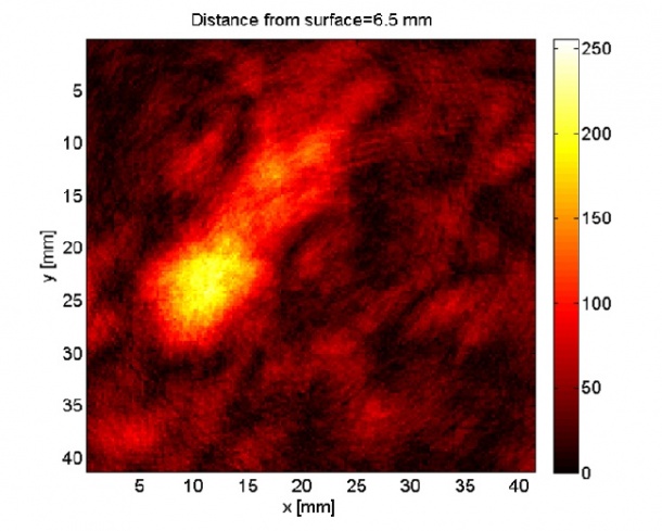
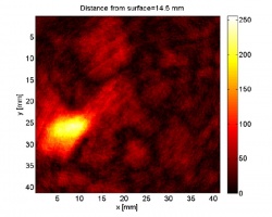
‘Photoacoustics can visualise angiogenic processes associated to tumour growth and its suppression by therapy. For instance, we focus on photoacoustic mammography for early detection and improved diagnosis. If tumours are to be followed during therapy, photoacoustics is attractive in terms of radiation load and probably also in terms of costs. A further sophistication of photoacoustics is spectroscopy, which may enable further diagnosis of tumours by their oxygen saturation. An interesting development is the possibility to provide anti-tumour drugs with an optical label. While this labelling is done with the purpose of fluorescent imaging, these labels can be visualised with photoacoustics too. This would turn photoacoustics into a tool for targeted therapy management. ‘A much less investigated application field of high relevance is rheumatology. Diseases such as osteoarthritis and rheumatoid arthritis affect a large number of people, and diagnosing these diseases at an early stage is difficult. In a recently granted European project, a range of partners including Osram, ESAOTE Europe, Quantel, and University of Twente, team up to realise a compact ultrasound-photoacoustic device for this purpose. ‘While (Doppler) ultrasound imaging can visualise synovial inflammation through the presence of big vessels, photoacoustics also targets the smaller vessels, potentially giving higher sensitivity for earlier signs of inflammation. A device like this will also be applicable for skin, such as skin cancer and ‘port wine’ stains.’
What will need to change in hospitals when photoacoustic imaging is embraced?
How easily photoacoustics is introduced into the existing workflow depends on the application. A compact hybrid photoacoustic-ultrasound imager is basically an ultrasound imager with a photoacoustic option. It may be a portable or bedside device that will be easily accepted and integrated in existing procedures. The position of photoacoustic mammography in the entire diagnostic chain may vary from completely replacing X-ray in screening, or being an additional tool in the complete chain, to a tool for monitoring breast-conserving therapies. It would be great if photoacoustics could lead to a reduction in unnecessary hospitalisations or if we could screen younger women than is currently the case with X-ray mammography.’
What will the cost be?
‘This is very difficult to predict. In addition to the purchase price of the device, logistics costs should be taken into account. In the end, what counts is the total healthcare benefit, to be determined by health technology assessment methods. As for the device price: for devices based on normal ultrasound scanners, the photoacoustic option will lead to a moderate cost increase if diode laser technology can be used. This is the aim of our new European project. However, for the optical mammoscope the device price will be higher, because a more powerful laser is needed and a specific tomographic detector arrangement has to be developed.’
When might photoacoustics become part of routine work?
‘For the introduction of photoacoustics as an integrated part of a normal scanner, I expect an introduction time of about five years. In addition to developing the device, clinical research must be performed aiming at developing medical applications. For mammography it’s rather a ten year period before the technology will have secured a position within breast cancer care.’
04.09.2012