Image by Bob Williams from Pixabay
News • Inhalation visualised
New imaging tech gives insights into pulmonary drug delivery
Inhalation therapy is widely used for the treatment of lung diseases. Targeting of drugs to the site of disease is a major goal to improve drug efficacy and minimize side effects.
Scientists at the Helmholtz Zentrum München and the Technical University of Munich have now shown that combined insight from various imaging methods allows for real-time monitoring of the dynamic process of drug delivery and high-resolution visualization of the pulmonary drug distribution in murine lungs. This new technology provides guidance on optimizing pulmonary delivery techniques and designing novel nanoparticle-based therapeutics. The paper was published in SMALL journal.
© Gradl & Pfeiffer, TUM
Optimizing drug delivery to the lung begins with understanding the dynamic process of drug delivery. While the underlying mechanisms are generally well known for aerosol inhalation, this is not the case for non-aerosol based methods, which are even more widely used in preclinical lung research. By visualizing the process of drug delivery in real-time, the team of researchers unveiled the specific mechanisms of drug delivery and their implications for drug distribution in a preclinical animal model. These findings open new avenues for optimization of pulmonary drug delivery in patients.
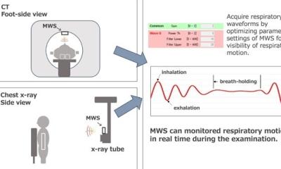
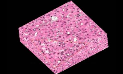
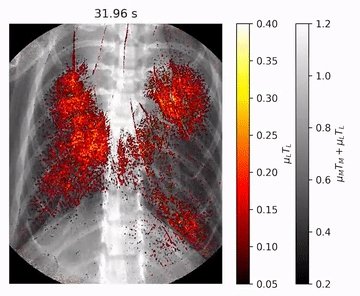
The imaging platform presented in this new technique consists of five preclinical imaging methods relying on propagation-based phase contrast X-ray imaging (PB-PCXI; two modes) and fluorescence imaging techniques (three modes). When delivering a mixture of nanoparticle-based contrast agents for X-ray and fluorescent imaging, the former can be used to visualize in real-time the dynamic delivery process in living mice, while the latter reveals the quantitative, three-dimensional drug/nanoparticle distribution profile at cellular resolution in ex vivo lungs.
The complementary information obtained from this imaging platform has revealed the previously unknown exact mechanisms of pulmonary drug delivery for intratracheal instillation and nasal aspiration. Moreover, it has revealed the reason for unexpected dose “hot-spots” in murine lungs after ventilator-assisted aerosol inhalation. With this technology novel concepts of targeting drugs to specific regions of the lung can be studied on live animals. It optimizes drug delivery techniques due to immediate information on the success (or failure) of novel drug delivery methods such as nanoparticle-based therapeutics. This is expected to expedite preclinical testing. After optimization in animal models these concepts will be translated into improved therapeutic outcome of inhalation therapy in patients.
This research demonstrates successful marriage of two cutting edge imaging technologies for preclinical studies of the lung, namely propagation-based phase contrast X-ray imaging (PB-PCXI) utilizing the Munich Compact Light Source (MuCLS) and light sheet fluorescence imaging (LSFM) which was established at the Munich School of BioEngineering at TUM lead by Prof. Franz Pfeiffer and the Helmholtz Zentrum München lead by Dr. Otmar Schmid, respectively. This research was partially supported through the EU Horizon 2020 project SmartNanoTox, grant agreement no. 686098
Source: Helmholtz Zentrum München
26.10.2019