Article • From threat to essential ally
Automation, not replacement: the true promise of AI in radiology
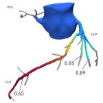
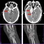
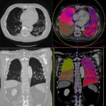
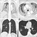
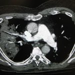
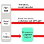
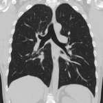
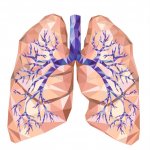
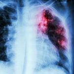
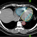
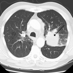
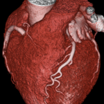
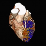
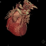
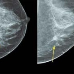
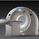
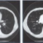
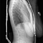
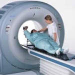
Will artificial intelligence (AI) render radiologists obsolete? What seemed a likely scenario only nine years ago, has now given way to a quite different reality: At RSNA 2025, two experts outlined how AI tools are becoming essential allies – not replacements – for radiologists facing an unprecedented workforce crisis.