Fast. Precise. Sharp.
Superb Microvascular Imaging and more…
Impacting on clinical decisions. Accelerating clinical routine. Following the release of its new Version 6 software upgrade for the Aplio Platinum Series ultrasound system, Toshiba has received high marks for the enhanced functions and performance from practitioners, each offering specific insights into how they are applying the technology.
In a whirlwind world tour, here’s what they say: Superb Microvascular Imaging
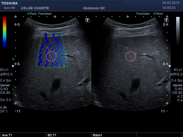
‘Estimated very conservatively, I’d say that in around 20% of cases a different approach to treatment results from the use of Superb Microvascular Imaging (SMI),’ reports Professor Thomas Fischer MD, Head of Ultrasound Diagnostics at the Institute for Radiology at the Charité Mitte Hospital in Berlin.
SMI is a Doppler imaging procedure that reacts a lot more sensitively to low flow speeds than normal Doppler imaging – with the added benefit of increased spatial and temporal resolution. The new version of the software has also reduced clutter artefacts that can affect the visualisation of perfusion. SMI can be used with contrast agents, can be visualised in 3-D and is compatible with more transducers.
In clinical diagnostics SMI has proved to be a game changer.
Fischer suggests this functionality can be used wherever the objective is the diagnosis of vascularisation and he sees a particular advantage for the diagnosis of liver disease and, specifically, cancers. After the wash-in and wash-out phases of the contrast agent, the examiner can switch to SMI, now optimised for use with contrast, and visualise vascular patterns, long after the bolus injection, he explained. ‘It is important for the detailed diagnosis to see how vessels grow into the tumour and to visualise exactly the vascular tree of the tumour, which is now also possible in 3-D technology with SMI.’
Meanwhile, half a world away at the Royal Melbourne Hospital in Australia, Robert Gibson MD finds Version 6 enhancement on the Aplio platform to be refined SMI and simplified its use with filter controls, which, he said, ‘should prove a real clinical advance in displaying small vessel flow and vascular morphology.’
Shear-wave elastography
An essential innovation of the Aplio Platinum series was the introduction of the shear-wave elastography with propagation mode, also making it possible to visualise the propagation of the shear-wave generated in the tissue as a colour-coded image, while simultaneously measuring the absolute value of elasticity in a chosen region.
‘Having these available in twin view allows a greater degree of confidence in selecting reliable regions of interest for elastography measurement,’ Gibson said. Elastography mapping in general has been enhanced with the Version 6 upgrades to the Aplio platform, he added, with propagation map cleaner than ever. ‘The Aplio 500 can indicate whether an elasticity measurement was successful, or not, because of the propagation mode, and the new version has even further improved this,’ Fischer said. ‘The examination procedure used to be a case of guessing the region of interest in a certain section of the image, starting the measurements and hoping that shear-wave signals were actually being measured. Now we can actually see whether or not the quality of the shear-wave propagation is adequate and then measure where propagation lines occur most evenly within the region of interest.’
‘This enhanced functionality carries clinical impact,’ he pointed out. ‘It will lead to long-term changes for the diagnosis of liver disease. Whilst it is possible to diagnose fibrosis with the help of a biopsy, shear-wave measurements can document much larger sections of the organ. To me this makes more sense than examining just a small sample.’
Fusion imaging and real-time 3-D needle tracking
Three rooms are equipped for interventional radiology at the University Hospital of Strasbourg in France and, according to the department chairman Afshin Gangi MD, ‘They are packed with patients, completely full, because we are asked to do so many interventions. ‘The majority of our cases are for biopsies, but patients are lined up for ablations, as well.’ When the upgraded needle-tracking capability arrived for the Aplio Platinum system, ‘it was like adding a fourth room for biopsies and interventions,’ he said. ‘Once I have the image fusion and know where the tumour is situated, I can do the intervention anywhere I can correctly position a patient. With the Toshiba Aplio platform, I don’t have to wait for the CT or the MR room to be open.’
Additionally, Gangi pointed out, ‘The needle-tracking function is excellent, giving an ability to truly project the path for the needle into the lesion, and then keeping operating clinicians on-track, even in places where the actual needle cannot be visualised by ultrasound because of the bone structures or body fat.
‘The system shows exactly where the tip of that needle is and, even better, tells me if I am too high or too low. We can achieve far greater precision during biopsies now – more precise every time. With the help of image fusion and needle navigation we can now use ultrasound guidance for applications we would never have considered before, such as those of the bone or the chest wall.’
The ease-of-use and accuracy of the enhanced image fusion software has also affected clinical routine and accelerated patient throughput, Gangi explained: ‘Today the technicians can do this so that when the interventionalist comes into the room, it’s done. This saves a lot of time, and I can tell you this is very important for us.’
Toshiba is at Medica Hall 9/D05
16.11.2015