Article • MEDIRAD project
Increasing the protection from medical radiation
Important findings from a Europe-wide project to improve medical radiation protection for patients and staff were detailed at a session of the ECR in Vienna. The five-year MEDIRAD project sought to enhance the scientific basis and clinical practice of radiation protection in medicine by better understanding and evaluating the health effects of exposure to low-dose ionising radiation resulting from diagnostic and therapeutic procedures.
By Mark Nicholls
A multi-disciplinary consortium of various research groups, with 34 partners in 14 countries, looked at areas of dose evaluation and optimisation in medical imaging; the impact of low dose radiation exposure; breast radiotherapy and secondary cardiovascular risks; and the impact of paediatric scanning.
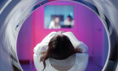
Patient protection

Image source: Barcelona Institute for Global Health
With MEDIRAD having made a number of recommendations for enhancing the effective protection of patients and medical professionals, as well as for identifying further research priorities, the ERC session heard speakers discuss new optimisation methods in chest CT, early cardiovascular changes after radiotherapy, and paediatric CTs and cancer.
MEDIRAD scientific co-ordinator Elisabeth Cardis focussed on CT exposure in childhood to help understand cancer risk estimation for children, retrospective reconstruction of doses, and long-term follow-up. ‘The reason we study paediatric exposure to ionising radiation is because there are concerns about potential health impacts of radiation exposure in childhood,’ said Professor Cardis from the Institute for Global Health in Barcelona. ‘Exposure at a young age is associated with a higher relative risk of several forms of cancer than exposure later in life.’
Risk estimates
Highlighting how the number of paediatric CTs has grown dramatically in high income countries in recent decades through improvements in technology and more applications for paediatric patients, she said the project concluded that paediatric CT scanning appears to increase the risk of cancer, particularly in haematological malignancies and brain tumours in a dose dependent fashion. She said the MEDIRAD preliminary findings were that missing doses from conventional x-rays could lead to an underestimation of risk in higher dose categories, though this needs to be confirmed in a full study. However, while final data from the study is still awaited, there appears ‘substantial improvements in precision of risk estimates.’
Cancer risk

Photo: supplied
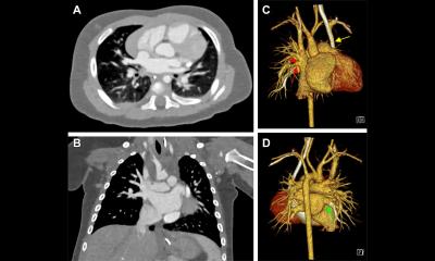
John Stratakis from the University of Crete’s Department of Medical Physics, discussed the element of patient-specific radiation dose and cancer risk estimation from chest CT. He also detailed a novel CT dosimetry tool (CTRAD) developed within the framework of the MEDIRAD project.
Dr Stratakis focussed on understanding the limitations of current CT dosimetry methods and tools, the need to appreciate the value of accurate patient dosimetry in CT and the value of patient-specific methodologies for calculating doses and risks in CT. ‘CT dosimetry has not escalated in pace with the exponential development of CT technology,’ he said. Various alternatives for more accurate CT dosimetry have been proposed over the last two decades, but he suggested they have limitations.
High accuracy
The expert said a high level of accuracy is needed in CT dose measurements because there is evidence of non-cancer effects such as lens opacities, cataracts and cardiovascular disease at relatively low doses; while patients subjected to multiple CT scans and exposed to doses higher than the 100mSv threshold are at significant stochastic risk. He said: ‘Inaccurate patient dosimetry can underestimate or overestimate the radiation burden associated with the recurrent CT imaging, leading to wrong priorities and incorrect dose management of these patients.
‘We actually need accuracy with dosimetry because accurate dose estimation is the pillar of exposure justification, protocol optimisation, and is invaluable to assess organ radiation doses and radiation use risk of patients. It conclusively governs the management of patients undergoing CT procedures.’
CTRAD tool
He underlined the value of personalized CT dosimetry methods, where a scanner-specific and patient-specific organ-dose estimation is based on a procedure that combines computational techniques and patient CT scans via the CTRAD image-based calculation tool. A free tool, autoWED, included in CTRAD to automatically calculate Water Equivalent Diameter (WED), enables the correlation of organ dose to patient attenuation characteristics.
Dr Stratakis explained that with CTRAD, users can upload a single DICOM image or a DICOM series for dosimetric calculations. CTRAD, he added, can calculate risk and a printable report can provide a summary of the absorbed dose of primarily-irradiated organs as well as the life attributable risks for cancer incidence and mortality. He said the tool has future-proofed characteristics with elements of sustainability, maintainability, reliability, and security; is web-based; and it is easily upgradable with additional data for scanners and anatomical regions.
Personalized dosimetry
AI has the future potential to accelerate computations and segmentations to eliminate limitations and provide organ dose estimates in real time
John Stratakis
In conclusion, he said: ‘Current CT dosimetry based on simple metrics may have weaknesses, so gold standard combined with Monte Carlo simulations personalized dosimetry can bring a paradigm shift in CT dosimetry, providing accurate organ dose estimates. ‘AI has the future potential to accelerate computations and segmentations to eliminate limitations and provide organ dose estimates in real time.’
The session also heard presentations on other elements of the MEDIRAD project looking at cardiac CT and cardiac MRI biomarkers before and after breast cancer irradiation; effectiveness of staff radioprotective equipment during interventional procedures; and how MEDIRAD results can be translated into daily clinical practice.
Profiles:
Elisabeth Cardis is a research professor in radiation epidemiology at the Institute for Global Health in Barcelona and MEDIRAD scientific coordinator. She is the author of over 100 indexed publications and a member of the board of the MELODI (Multidisciplinary European Low Dose Initiative) platform.
John Stratakis is a research associate in the University of Crete’s Department of Medical Physics and a medical physicist in the University Hospital in Heraklion, Crete.
21.07.2022