Image credit: Kindai University
News • Successful treatment of congenital heart disease
First “Double-decker” surgery for scimitar syndrome
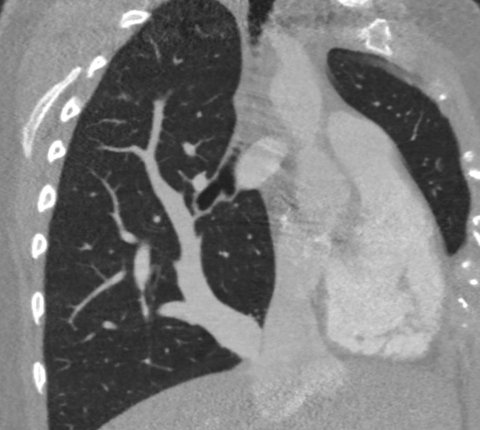
Scimitar syndrome, a rare congenital heart disease, involves an anomalous pulmonary venous return where the right pulmonary veins return to the inferior vena cava instead of the left atrium.
It is mainly diagnosed in infants, with an estimated prevalence of 1–3 per 100,000 births. Delayed treatment can lead to pulmonary hypertension, right heart failure, respiratory failure, heart arrhythmia, and growth disorders.

Image source: Matthew Cham, MD, Scimitar syndrome chest CT (CC BY-SA 4.0)
This syndrome is characterized by anomalous pulmonary venous drainage to the inferior vena cava, and the usual surgical repair involves re-implanting the right pulmonary veins (scimitar vein) to the left atrium or creating an intra-atrial tunnel to redirect the scimitar vein to the left atrium. However, these methods have a critical problem of postoperative pulmonary vein obstruction. If this occurs, it can lead to severe pulmonary venous congestion and subsequent hemoptysis. In such patients, the success rate of re-intervention for pulmonary venous obstruction is very low.
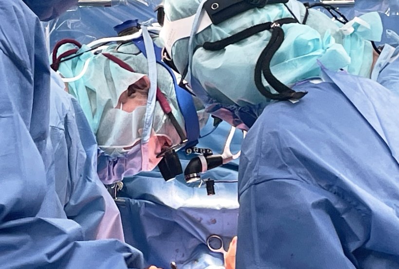
Surgeons at Kindai University Hospital have just performed an unprecedented surgical procedure on a two-year-old child diagnosed with scimitar syndrome. The procedure was the world’s first successful application of the “Double-decker Technique” used to repair another type of partial anomalous pulmonary venous return. This new procedure, a modified double-decker technique for scimitar syndrome, uses no artificial material and reconstructs two blood flow pathways (right pulmonary vein and inferior vena cava) using only the patient’s atrial wall. This novel surgery was conducted by a pediatric cardiac surgery team led by Senior Professor Genichi Sakaguchi, Affiliate Professor Shinichiro Oda, and Associate Professor Satoshi Asada from the Department of Cardiovascular Surgery, Kindai University Hospital.
The patient was referred to the hospital for suspected congenital heart disease from another public hospital after developing a fever. The patient was diagnosed with scimitar syndrome by cardiac echocardiogram and contrast-enhanced computed tomography. After the surgery, there were no problems with the reconstructed blood flow pathways in the right pulmonary veins and inferior vena cava. The patient was discharged from our hospital 10 days after the surgery without any postoperative complications.
The advantage of this new technique is that the new blood flow pathway of the inferior vena cava is created outside (on the pulmonary venous pathway), the team reports. This technique can create wide pathways separately and reduces the risk of obstruction. The conventional intra-atrial tunneling divides the inferior vena cava into two pathways for the right pulmonary vein and the inferior vena cava. That is why the conventional technique is likely to create narrower pathways and develop obstructions. In addition, the surgical site is expected to grow following the patient’s somatic growth because these pathways were reconstructed by the pedicled atrial wall without any artificial material.
The success of this surgery can make this a common surgical technique for scimitar syndrome, and surgical outcomes for this rare disease are expected to be improved in the future, the team concludes.
Source: Kindai University
22.07.2024


