Credit: American Journal of Roentgenology (AJR)
News • What radiologists need to know
How lung disorders like COVID-19 affect children
Although the clinical symptoms of new pediatric lung disorders such as severe acute respiratory syndrome (SARS), swine-origin influenza A (H1N1), Middle East respiratory syndrome (MERS), e-cigarette or vaping product use-associated lung injury (EVALI), and coronavirus disease (COVID-19) pneumonia may be nonspecific, some characteristic imaging findings "have emerged or are currently emerging," according to an open-access article in the American Journal of Roentgenology (AJR).
Credit: American Journal of Roentgenology (AJR)
"Although there are some overlapping imaging features of these disorders," wrote first author Alexandra M. Foust of Boston Children's Hospital and Harvard Medical School, "careful evaluation of the distribution, lung zone preference, and symmetry of the abnormalities with an eye for a few unique differentiating imaging features, such as the halo sign seen in COVID-19 and subpleural sparing and the atoll sign seen in EVALI, can allow the radiologist to offer a narrower differential diagnosis in pediatric patients, leading to optimal patient care."
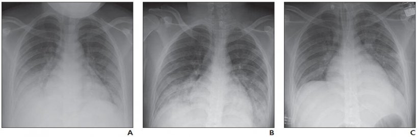
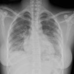
At most institutions, whereas the first imaging study performed in patients with clinically suspected COVID-19 is chest radiography, Foust and colleagues' review of the clinical literature found that studies on chest radiography findings in patients with COVID-19 were relatively scarce. Regarding the limited studies of pediatric patients with COVID-19, Foust et al. noted chest radiography "may show normal findings; patchy bilateral ground-glass opacity (GGO), consolidation, or both; peripheral and lower lung zone predominance."
Recommended article
Interview • Chest X-ray, CT and more
Imaging the coronavirus disease COVID-19
Chest X-ray is the first imaging method to diagnose COVID-19 coronavirus infection in Spain, but in the light of new evidence this may change soon, according to Milagros Martí de Gracia, Vice President of the Spanish Society of Radiology (SERAM) and head of the emergency radiology unit at La Paz Hospital in Madrid, one of the hot spots for viral re-production of COVID-19.
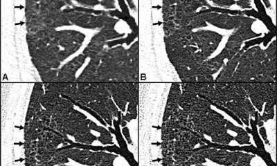
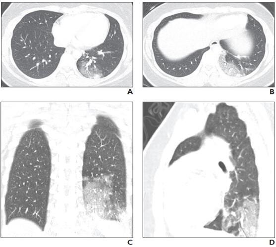
Similarly, while the literature describing chest CT findings in patients with COVID-19 are more robust than those describing chest radiography findings, only a few articles have reported CT findings of COVID-19 in children. A study of 20 pediatric patients with COVID-19 reported that the most frequently observed abnormalities on CT were subpleural lesions (100% of patients), unilateral (30%) or bilateral (50%) pulmonary lesions, GGO (60%), and consolidation with a rim of GGO surrounding it, also known as the halo sign (50%).
The authors of this AJR article also pointed to a smaller study of five pediatric patients with COVID-19, where investigators reported modest patchy GGO, one with peripheral subpleural involvement, in three patients that resolved on follow-up CT examination.
Source: American Roentgen Ray Society
11.05.2020