IGS
The GPS of arteries
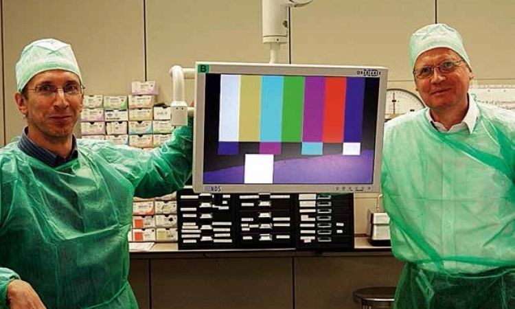
In our last issue we featured the Future Operating Room Project developed at St Olavs Hospital, University Hospital of Trondheim, Norway, a collaboration between the hospital and the Norwegian University of Science and Technology. There, highly promising research on navigation is being carried out in co-peration with the research foundation Sintef Health Research. Professor of Surgery Hans O Myhre (above) of St Olavs and Senior Research Scientist Jon Harald Kaspersen PhD (below), of Sintef, describe the project's aims, their development and clinical testing of the navigation system CustusX, and the need that has now arisen for industrial collaborators to take the team's discoveries forward to expand indications for endovascular therapy in general
Image-guided surgery (IGS) is based on an evolving technology, and is mainly developed for minimally invasive procedures. Minimally invasive treatment has expanded tremendously during recent years. It represents a decreased trauma to the patient compared with open surgery. This could again lead to shorter convalescence and hospital stay and less postoperative pain for the patient.
So far, the majority of developed systems for IGS have been designed for neuro-surgery, but other clinical applications are emerging, such as otologic procedures, hepatic surgery, orthopaedic surgery and endovascular procedures. In the most advanced systems available today, IGS provides the surgeon with a two- (2D) and three-dimensional (3D) visual ‘road map’ of the patient’s anatomy, including the surgical instruments used during the procedure. This is similar to the situation where a ship with a GPS (global positioning system) installed can plot its position on a map. Surgical navigation depends on a ‘map’ of the body, such as a CT-volume combined with information about where the surgeon’s tool is located. The latter is provided by a positioning system. Prior to the development of IGS, surgeons performing minimally invasive surgery (MIS) could only see the surface area visible from the end of the imaging device, e.g. a laparoscope. IGS overcomes this limitation and provides the surgeon with real-time enhanced visualisation. Thereby the surgeon can gain access to anatomic areas that otherwise would be difficult to reach.
An IGS system combines a high-speed computer system, specialised software and tracking technology. On this computerised system the actual movements of the surgical instruments are correlated with the patient’s preoperative medical images and are displayed on the system’s monitor. The precision of computerised instrument localisation and navigation is critical for manoeuvring safely within concealed anatomy and for the surgeon to perform more precise and careful surgery. However, most IGS systems are based on preoperative images, sometimes acquired the day before surgery. This means that, as surgery proceeds, the images could become continuously less representative for the actual anatomy of the patient. Introducing intra-operative imaging modalities, such as ultrasound, can solve this problem.
Prior to surgery, the MR/CT images are imported into our in-house developed navigation software, CustusX, and reconstructed into a regular 3-D volume. The preoperative MR/CT images are registered to the patient. Surfaces of a few essential organs are extracted from the image data and visualised in a 3-D scene. The surgical tool controls the visualisations on the navigation monitor, and these images are updated continuously (in real time) according to both position and orientation of the pointer.
In addition it is possible to distribute the 3-D intra-operative scene to the radiology department using CustusX. Both the radiologist and the surgeon in the operating room are able to interact with the data. With this collaborative feature it is possible for the surgeon to get expert advice without having the radiologist present in the operating room.
Endovascular therapy
Endovascular techniques are increasingly used in the treatment of diseases of the main blood vessel (aorta) in the chest or abdomen. Previously, it was necessary in all cases to use large incisions and these operations were more traumatic, necessitating a longer stay in the intensive care unit (ICU) and the ward. Following minimally invasive surgery e.g. of an abdominal aortic aneurysm, the patient can get out of bed after a few hours and prepare his own breakfast the next morning.
However, so far some patients cannot be treated by endovascular technique because the disease is rather extensive without a place for fixation of the stentgraft. A stentgraft is a synthetic prosthesis consisting of a textile part with a thin metal net on the inside. The stentgraft is compressed and delivered into an introducer system. This implant is introduced into the vascular system through a small incision in the femoral artery in the groin and regional anaesthesia is applied. As soon as the proper position for the stentgraft is obtained the introducer system is withdrawn and the stentgraft expands. It is fixed to the arterial wall by radial forces, hooks and barbs. The patient is awake during the procedure. In most cases this technique is used for aortic aneurysmal disease (dilatation of the aorta) or so-called aortic dissection where the blood is running in a false lumen within the arterial wall.
In order to offer endovascular techniques to a broader group of patients, there is a need for stentgrafts with side branches or fenestrations, which can lead the blood from the main prosthesis into the major arterial branches to the head, arms, kidneys, liver and bowel. To obtain this there is need for a new prosthesis, as mentioned. Furthermore, we need flexible introducer systems and the stentgrafts with the side branches need to be deployed accurately and easily.
The surgical tools used in endovascular therapy are guidewires and catheters. Therefore, navigation will depend on well-functioning micro-positioning sensors, which can be adapted to these small instruments. We also think it is possible to adapt micro-positioning sensors (<1mm) to the stentgraft itself. We are looking for industrial collaborators who can combine the small sensor with catheters, introducers or perhaps stentgrafts, and think that this is going to expand the indications for endovascular therapy in general. For navigation our own system CustusX meets these requirements.
We are also using the so-called DYNA CT principle (Siemens) where the C-arm of an angiography unit rotates providing us with real-time 3D-images of the vascular tree. With these techniques we think that within a few years we will be able to repair most parts of the arterial system using minimally invasive technique.
The navigation system could also be useful for re-operations where arteries and other structures are embedded in heavy scar tissue and where dissection is difficult. Development and clinical testing of CustusX will be one of the main activities within the endovascular Future Operating Room project at St. Olavs Hospital in Trondheim.
Contacts: hans.myhre@ntnu.no
jon.h.kaspersen@sintef.no
Details: www.stolav.no/FOR
www.sintef.no/Medtech
03.08.2006