Source: Fujitsu
News • Joint Research
AI helps diagnosing Covid-19
Fujitsu and Tokyo Shinagawa Hospital today announced the launch of a joint R&D project for AI technology to support diagnostic imaging via chest CT (Computed Tomography), which represents a promising candidate for the effective diagnosis of COVID-19 pneumonia.
Fujitsu and Tokyo Shinagawa Hospital anticipate that the system will deliver early detection of cases of Covid-19 pneumonia based on chest CT image findings, even in cases in which the possibility of infection is determined to be low upon initial examination. This joint research will enhance AI diagnostic support technology for novel coronavirus pneumonia, and Fujitsu ultimately aims to commercialize the technology as a healthcare solution for frontline medical professionals.
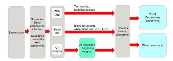
When treating patients who are strongly suspected of having a novel coronavirus infection, the diagnosis and treatment plan are determined holistically based on PCR test results as well as other test results including blood tests and chest CT test. Even if the PCR test is negative, the diagnosis of new-onset coronavirus pneumonia can be made by other tests, and imaging with chest CT is an important consideration. Doctors make lung disease diagnoses based on the characteristics of the shadows in the lesion on the patient's chest CT image. In addition to identifying abnormalities, they visually check hundreds of chest CT images per patient to understand the three-dimensional distribution of shadows throughout the lung. A growing demand for technologies that reduce the burden on doctors and support speedy decision-making has accompanied the spread of the Covid-19 virus. It is also important to identify novel coronavirus pneumonia from a CT scan of the patient's chest if there is a low likelihood of a novel coronavirus infection during the examination and PCR testing is not performed.
In response to the need for diagnostic support, Fujitsu and Tokyo Shinagawa Hospital will jointly embark on the development of AI-assisted diagnostic imaging technology for chest CT scans, which are considered to be an effective method in diagnosing novel coronavirus pneumonia.
Past CT image data of Covid-19 pneumonia patients from Tokyo Shinagawa Hospital will aid in the development of AI technology that detects abnormal shadow patterns in the lungs. As the AI learns from this data about the possibility of Covid-19 pneumonia, Fujitsu and Tokyo Shinagawa Hospital will work together to evaluate the effectiveness of the technology. When diagnosing Covid-19 pneumonia, patterns of abnormal opacities in the lungs as well as the spread of shadows across the entire lung is important information.
Recommended article

News • Reducing coronavirus test burden
AI speeds up COVID-19 screening in emergency rooms
Researchers from Eindhoven University of Technology (TU/e) and the Catharina Hospital in Eindhoven have developed a new algorithm for the rapid screening for COVID-19. The software is intended for use in Emergency Rooms (ER), to quickly exclude the presence of corona in incoming patients. As a result, doctors need to conduct fewer standard coronavirus tests, increasing efficiency. The quick scan…
Patterns of abnormal shadows are detected using AI developed by Fujitsu Laboratories, Ltd. The lung is divided into four areas on the CT image: the right lung periphery, the right lung center, the left lung center, and the left lung periphery. The distribution of shadows in the vertical direction in each area is displayed in a histogram. This will enable the development of new AI that can quantify the three-dimensional spread of shadows, while using the detected abnormal shadow patterns and shadow distributions to identify Covid-19 pneumonia.
While showing the possibility of Covid-19 infection with the AI technology, the aim is to shorten the amount of time doctors spend visually confirming the three-dimensional spread of the shadow from hundreds of chest CT images and to allow even non-specialists to efficiently diagnose Covid-19 pneumonia.
Through this joint research and development, Fujitsu and Tokyo Shinagawa Hospital aim to deliver technology that leverages a variety of information for diagnosis of Covid-19 pneumonia to achieve improved diagnostic support from chest CT image diagnosis. Fujitsu is considering the commercialization of this technology as a healthcare solution. By linking it with electronic medical record information, the goal is to support diagnosis by doctors based on chest CT images, while also expanding the application area of this technology. Tokyo Shinagawa Hospital aims to contribute to society by integrating various studies conducted in the hospital and using them for diagnosis and treatment of novel coronavirus pneumonia.
Source: Fujitsu
03.09.2020