

News • Rad Companion
Siemens expands AI portfolio in clinical decision-making
The AI-Rad Companion family supports radiologists, radiation oncologists, radiotherapists and medical physicists through automated post processing of MRI, CT and X-ray datasets. It saves the clinicians' time and helps them to increase their diagnostic precision.
The steady rise of radiology examinations and staff shortages lead to a limited amount of time per case as well as an increasing danger of missing clinically relevant findings. Siemens Healthineers is continuously working on further solutions to expand and enhance its AI-Rad Companion algorithms. These algorithms are trained, tested and validated against a substantial amount of data sets. The data sets are selected to reflect the diversity of the population, avoid bias and deliver reliable results.
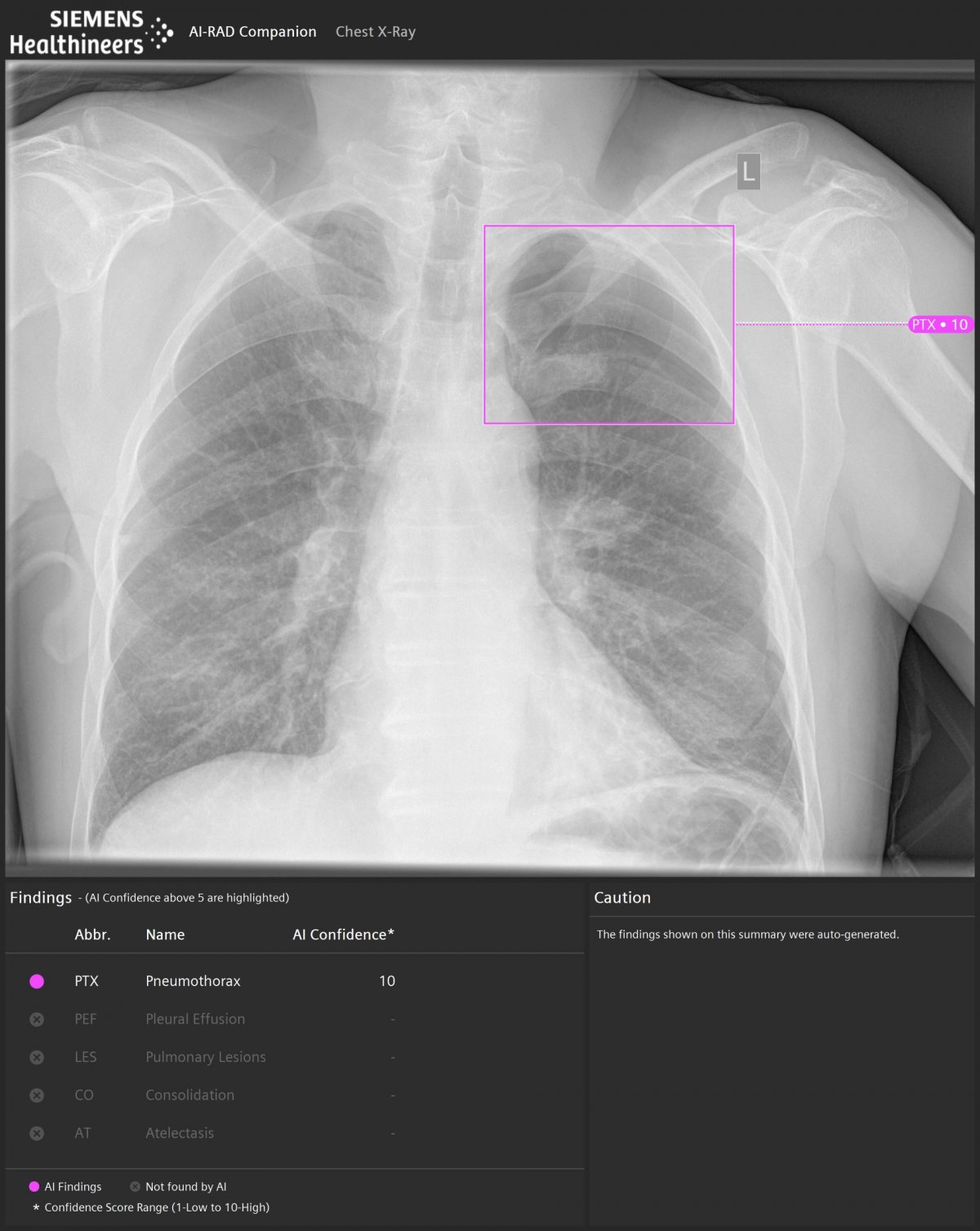
Image courtesy of University Hospital Munich, Munich, Germany
The new CE-labeled AI-Rad Companion Chest X-ray1 helps to detect radiographic findings on upright Chest X-ray images from multiple vendors2 and acts as a companion for the clinician. Its algorithms detect and characterize several findings in the lung and pleura such as pulmonary lesions, pneumothorax, pleural effusion, consolidation and atelectasis and indicate their probability with confidence scores. AI-Rad Companion Chest X-ray supports the DICOM format and delivers PACS ready results while reading the original image. “AI-Rad Companion Chest X-ray is fully embedded in the reading workflow and the representation of outcomes is presented in a perfect manner. It delivers the right information in a short and crisp format.”, reports Dr. Karsten Ridder, MVZ Prof. Dr. Uhlenbrock und Partner, Dortmund, Germany.
AI-Rad Companion not only helps to improve diagnostic accuracy, it also supports in increasing efficiency in Radiation Therapy Planning. The new AI-Rad Companion Organs RT3, CE-labelled, helps to handle the time-consuming manual contouring in multiple CT slice images, essential for Radiation Therapy Planning. Its AI algorithms help to achieve consistent high-quality contours through automated organs at risk contouring. “The use of AI-Rad Companion Organs RT makes our life easier. Especially the contouring of organs in the upper abdomen, leads to a noteworthy reduction of turnaround time.”, states Dr. Alexandros Papachristofilou at University Hospital Basel, Switzerland.
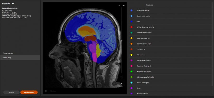
AI-Rad Companion also assists in the interpretation of MR images of the brain. The assessment of different brain areas is not easy. The FDA cleared and CE-marked AI-Rad Companion Brain MR4 for morphometry analysis automatically segments different brain areas and quantifies each of their volumes. These results are compared to a normative database and can easily be assessed by the radiologist through a color-coded deviation map and a quantitative overview. “From a clinical point of view, deviation maps are the most relevant for us. We primarily focus on them to support clinical diagnosis of dementia subtypes based on imaging phenotypes”, highlights Dr. Máté Maros, University Medical Center, Mannheim, Germany. The AI-Rad Companion portfolio includes further offerings that use MRI images for AI-based image quantification. AI-Rad Companion Prostate MR3 for biopsy support automatically segments the prostate and allows the manual annotations of lesions, which can support in targeted biopsies under MRI and Ultrasound fusion imaging. Targeted, MRI-supported biopsies like this can make it easier for the urologist to detect significant prostate carcinomas and improve the quality of patient care.
© Siemens Healthineers
The first member of the AI-Rad Companion family was launched in 2018. The AI-Rad Companion Chest CT offers a bundle of algorithms that help measure, highlight and segment relevant anatomies and abnormalities of thoracic CT images of the lung, heart, vertebrae and the aorta – all of them FDA-cleared and CE marked. The software automatically creates a standardized and quantitative report and sends its result to the reporting environment. “We processed 50 previously read studies as soon as we received AI-Rad Companion Chest CT. In 14 percent of the cases that were analyzed by the software, it delivered additional clinical valuable information that has not been noticed by the initial reading of the images”, states Dr. Ernesto Barrientos Manrique, Health Time Medica, Spain. “The findings in the remaining 86 percent of processed cases, were in line with the original reports submitted by our radiologists.”
All solutions in the AI-Rad Companion family are connected to the teamplay digital health platform. Applications in the cloud ensure that the customers’ clinical cases are processed in a secured and stable way. Moreover, customers easily receive the latest up-to-date algorithms via the platform and installation efforts are minimal.
References
- AI-Rad Companion Chest X-ray is currently under development; it is not for sale in the United States and other countries.
- Tested and validated on data from Siemens Healthineers, GE and Philips devices
- AI-Rad Companion Organs RT and Prostate MR are 510(k) pending and are not yet commercially available in the United States.
- AI-Rad Companion Brain MR is not commercially available in all countries, and its future availability cannot be ensured.
Source: Siemens Healthineers
16.07.2020


