Image source: RSNA
News • Imaging immunocompromised patients
Pneumonia diagnosis: deep learning takes the grain out of ultra-low dose CT
Denoised ultra-low dose CT can effectively diagnose pneumonia in immunocompromised patients using only 2% of the radiation dose of standard CT.
This is according to a study published in Radiology: Cardiothoracic Imaging, a journal of the Radiological Society of North America (RSNA).
“For patients with weakened immune systems, lung infections can be life threatening,” said lead study author Maximiliano Klug, M.D., a radiologist in the division of diagnostic imaging at the Sheba Medical Center in Ramat Gan, Israel. “CT scans are the gold standard for detecting pneumonia, but repeated scans can expose patients to significant radiation.”
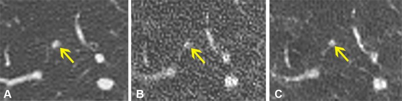
Image source: RSNA
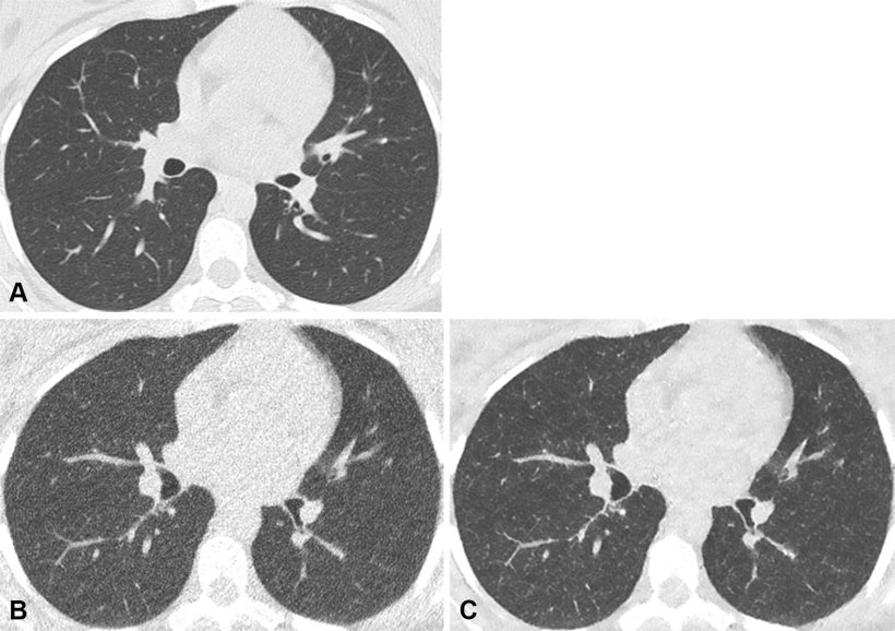
While the early diagnosis of lung infections in immunocompromised patients is important, the risks of cumulative radiation dose exposure from frequent CT scans is a concern. Ultra-low dose CT reduces radiation exposure but can result in poor image quality due to added “noise,” which manifests as a grainy texture throughout the image. This reduction in image quality can affect the accuracy of diagnosis. Therefore, Dr. Klug and colleagues sought to test the denoising capabilities of a deep learning algorithm on ultra-low dose CT scans.
From September 2020 to December 2022, 54 immunocompromised patients with fevers were referred to Dr. Klug’s division to undergo two chest CT scans: a normal dose scan and an ultra-low dose scan. A deep learning algorithm was applied to denoise all 54 of the ultra-low dose CT scans. Radiologists individually assessed and documented their findings from the normal dose CT, ultra-low dose CT and denoised ultra-low dose CT scans. They were blinded to all patient clinical information.
This study paves the way for safer, AI-driven imaging that reduces radiation exposure while preserving diagnostic accuracy
Maximiliano Klug
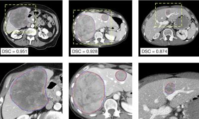
The deep learning algorithm significantly improved the image quality and clarity of the ultra-low dose CT scans and reduced false positives. Nodules were also more easily identified on the denoised scans. The average effective radiation dose for ultra-low dose scans was 2% of the average effective radiation dose of the standard CT scans. “This study paves the way for safer, AI-driven imaging that reduces radiation exposure while preserving diagnostic accuracy,” Dr. Klug said.
The researchers note that deep learning-based denoising on ultra-low dose CT scans can be beneficial in other patient groups, such as young patients.“This pilot study identified infection with a fraction of the radiation dose,” Dr. Klug said. “This approach could drive larger studies and ultimately reshape clinical guidelines, making denoised ultra-low dose CT the new standard for young immunocompromised patients.”
Future studies with larger sample sizes will help validate the findings from this study.
Source: Radiological Society of North America
14.03.2025