Article • The impact of AI
Radiology and radiologists: a painful divorce
Artificial intelligence based applications will replace radiologists in some areas, the physicist Bram van Ginneken predicts.
Report: Michael Krassnitzer
‘The profession of radiologist will change profoundly,’ predicts Bram van Ginneken, Professor of Medical Image Analysis at Radboud University Medical Centre. The cause is automatic image analysis by computers (first published in a paper in 1963) and deep learning, the method with which a computer learns to analyse images not by features extracted by a radiologist, but directly from the images themselves. The title of his talk during the European Congress of Radiology (ECR 2018) in Vienna speaks volumes: ‘Artificial intelligence and radiology: a perfect match. Radiology and radiologists: a painful divorce?’
For 50 years scientists had tried to build a system that could automatically analyse medical images. They failed. Humans are better. However, in 2013 they discovered that deep learning, an old idea from the 1970s, suddenly worked, thanks to better computers, more data to train on and improved algorithms to ‘educate’ artificial neural networks (the basis of deep learning). Currently, anything a human can do in one second, deep learning can also do. ‘Take any classification or detection task for which a human expert looks at an image for one second, or scrolls through a scan (and prior scan) for a few seconds, and then knows the answers – that task can also be done by a computer using deep learning at, or above, the level of the human expert. It’s still a lot of work to collect a well-annotated large data set and to engineer the deep learning system to perform well – but, it can be done.’
AI-based applications
In Ginneken’s view there are three types of applications: those replacing a radiologist’s task, those helping the radiologist with a task, and those doing something a radiologist does not do today – and probably never will. The application BoneXpert automatically measures bone age from a child’s hand X-ray, thereby replacing a human. ‘This program delivers a precise and standardised reading, so that 50 percent of radiologists no longer look at the images’, the Dutch physicist points out.
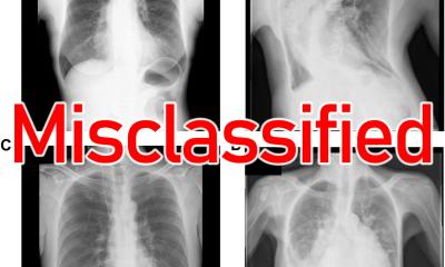
Detecting TB in X-rays
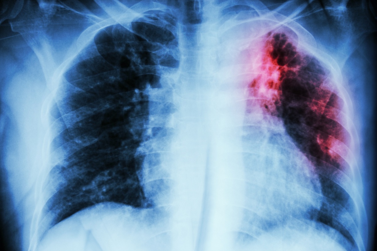
Another application of this type, CAD4TB (Computer-Aided Detect-ion for Tuberculosis) was co-developed by Ginneken. This is a software designed to help (non-expert) readers detect tuberculosis in chest X-rays. ‘The first version was released in 2011 and immediately used in Zambia, even before it was approved,’ Ginneken says. Meanwhile, CAD4TB is used in 24 other poor countries where radiologists are in short supply.
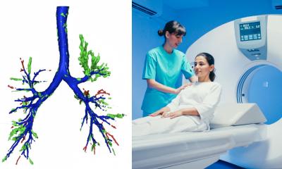
An example for an AI-based application assisting radiologists is Veolity, a lung screening workstation to read chest CT images efficiently. It includes automatic detection of nodules, automatic propagation of nodules found on prior scans, volumetric segmentation of solid, non-solid and part-solid nodules, with a single click, automatic lobe assignment and nodule type assessment, emphysema scoring and coronary calcium scoring. ‘With this software, radiologists are about 43 percent faster,’ Ginneken explains.
An example of the third type of app is StratX, a cloud-based quantitative CT analysis service that supports endobronchial valve (EBV) patient selection and therapy choice by providing clinically validated information on emphysema destruction, fissure completeness and lobar volumes. This system enables assessment of all potentially suitable patients for that minimally-invasive treatment for severe emphysema. ‘Radiologists have never done that,’ Ginneken points out.
Should physicians take over?
In terms of these automated services a question arises: Why involve a radiologist at all? Why don’t the treating physicians take it on? Ginneken mentions optical coherence tomography (OCT), an ophthalmology imaging technique that gives a very detailed view of the retina. The technique was developed by ophthalmologists and is used exclusively by ophthalmologists; radiologists were never involved. ‘I think this will be the case in any area of medicine where new imaging modalities are introduced.’ That situation does not sadden Ginneken. ‘Don’t think about radiology or jobs for radiologists,’ he says. ‘Think about the positive impact AI intelligence will have on healthcare, globally.’
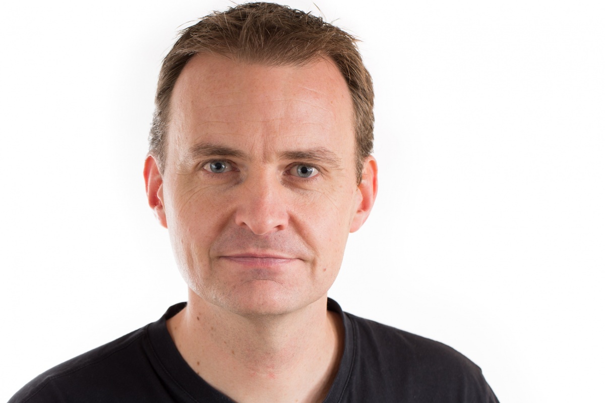
Profile:
Bram van Ginneken PhD is Professor of Medical Image Analysis at Radboud University Medical Centre and has co-chaired the Diagnostic Image Analysis Group since 2010. He also works for Fraunhofer Institute for Medical Image Computing (MEVIS) in Bremen, Germany, and is a founder of Thirona, a company that develops software and provides services for medical image analysis. Bram studied Physics at Eindhoven University of Technology and Utrecht University. In 2001, he gained his doc-torate at the Image Sciences Institute on Computer-Aided Diagnosis in Chest Radiography, and he has (co-) authored close to 200 publications in leading international journals.
07.05.2018