How low can the dose be?
Michael Maher, Professor of Radiology at the University Collage, Cork, Ireland, produced an answer to during a GE Healthcare Lunch Symposium at the European Congress of Radiology this March. It is 1.2 millisievert – at least for abdominal CT scans of Crohn’s disease patients.
Typically, dose reduction causes an increase in noise and image artifacts. But GE’s ASiR solves this by subtracting noise, not merely masking it. As a result, ASiR delivers enhanced image quality by improved low contrast detectability (LCD) whilst preserving anatomical detail.
"Crohn’s disease patients are a perfect example of a group that we frequently see in the hospital -- it is at high risk of getting radiation-induced cancer due to all the CT examinations these patients undergo during their lifetime. The question we followed at Cork University Collage was how to help those people and how to lower the radiation dose per examination.
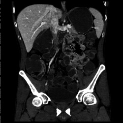
The average dose was 7.7 millisievert; studies reported that 5.6 millisievert were also possible. When we started our study with patients with Crohn’s disease, our ambition was to reduce the dose to the level of that of a regular film examination, which is 0.7 millisievert. Our solution for that ambitious aim was an iterative reconstruction software that enables us to get rid of the noise from the acquired pictures. The technology reduces the noise significantly and therefore improves the diagnostic acceptability of the images. With the iterative reconstruction technology we could reduce the dose to about 1.2 millisievert, which is very close to a regular film examination of the abdomen.
Our study includes approximately 40 patients and was prospective, so with those 40 patients we did two CT scans: one with 4.7 millisievert and one with ultra-low dose of 1.2 millisievert. First, we were not confident that 1.2 millisievert would be sufficient but, with the iterative reconstruction, we managed to get diagnostic images without compromising quality.
Of course, we are using the technology in all our patients but not pushing it to this level, as in the study, because we cannot do studies on every patient. But, to put it in a nutshell, we are finding that the technology is fantastic for doing CT scanning of the chest, brain and neck. It does result in improved diagnostic images at doses approximately 40% lower than without iterative reconstruction."
19.04.2011