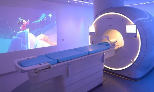
CT developments far excel those for MRI
For the sixth time, Alain Blum MD has invited the French CT community to Nancy to attend a symposium on multi-detector CT. The last invitations, two years back, drew several hundred radiologists and every CT manufacturer to Nancy for two days of debate, discussion and demonstrations.
‘There have been leaps in technology and developments that are shaking up clinical practice,’ explained Dr Blum, head of the radiology department at the University Hospital Centre in Nancy. ‘Every year we hear that CT is finished when, in fact, manufacturers continue to make enormous investments in research and each year has seen new developments far more significant than we have seen for MRI. Besides, this is also a personal thing – inviting people I like a lot.’
A third reason for organising the symposium, he told European Hospital, is that ‘Everyone in France is a bit depressed about the financial crisis that has impacted on the possibility for hospitals to acquire scanners. I think it becomes important to have a more optimistic discussion, telling colleagues we need to move forward, that we must not fall into a psychological morass, that technical developments are beneficial to everyone – the patient, radiologists, healthcare institutions,’ he explained. ‘We see, in some recent purchases, that people are ordering low-cost scanners to spend the least amount possible without taking into account that this will affect diagnostic quality and, after all, we need to maintain a sufficient level of quality.’
Endorsed by the French Society of Radiology, this year’s symposium was jointly organised by Dr Blum and Marc Zins MD, who leads the radiology group at Saint Joseph Hospital in Paris.
Innovations affecting diagnostic strategies and practice will be a focus for discussions with presentations on dual-energy image acquisition, spectral imaging, perfusion, iterative reconstruction and metal artefact reduction (MAR). Additionally, back end operation for image processing, structured reporting, or archiving and storage will be highlighted.
Managing radiation and dose will fill a whole session, as 2014 sees hospitals across France deploying new software for tracking and monitoring patient and staff exposure.
In Manufacturer’s Corner every CT-scan vendor will present products and the symposium will close with its signature final session featuring a face-off of consoles in a real-time demonstration.
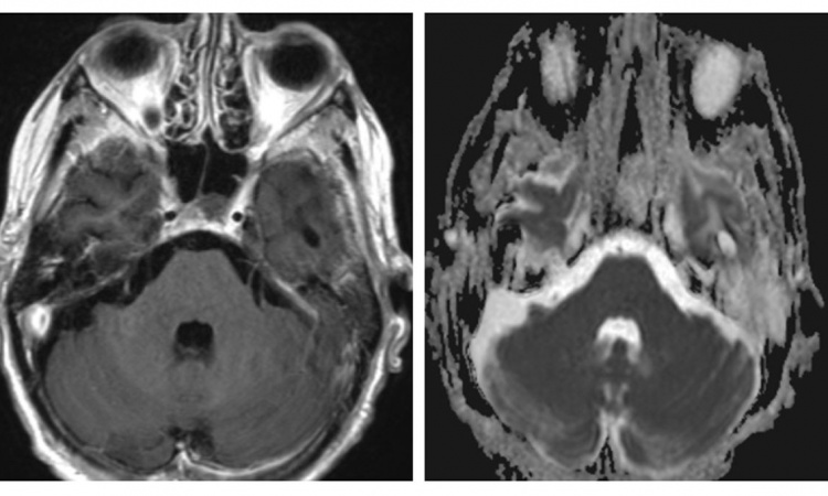
In charge of departments for musculoskeletal, emergency and neck radiology, it is not surprising that Dr Blum singles out advances in MAR as among symposium highlights. ‘Today all manufacturers offer with different levels of effectiveness a reduction in metal artefacts,’ he said. ‘This completely changes orthopaedic imaging. Up to this point we have been troubled with all the metal hardware, and now it is no longer a real obstacle. For example, with hip implants standard radiology shows the prosthesis and bones while the scanner can now show the implant, and show the bones, cement and affected soft tissue better. This becomes important taking into account that patients with pain associated with a prosthesis sometimes present with articular and peri-articular pseudo-tumours linked to a reaction that can be inflammatory or allergic.’
In other practice areas, the increasingly rapid speed of scanners has created new opportunities, notably for cardiac investigations, he added. This has come alongside reductions in radiation dose, and today effectively all CT exams are performed at lower dose. ‘Thanks to algorithms for iterative reconstruction, there have been great advances, and we will see this go even further with coming improvements,’ he said. ‘We’ve seen improvements in post-processing of image that’s much faster and efficient, which improves workflow.
‘We can also do thoracic scans at very low dose, which is why I invited two specialist to debate this around the question ‘Can We Leave Behind Thoracic Radiography?’ In other words, with a dose equivalent to traditional chest X-rays, can we do better with a scanner?’
‘In my opinion, we will see a migration from standard radiology to the CT scanner,’ he predicts. Today we do a lot of traditional chest X-rays because there is a good spatial resolution and it’s performed at a low dose; but a segmented scan is much more effective and, if today we can perform a scanner exam at the same dose as a chest X-ray, and can improve the workflow, then I believe we will progressively see a shift to thoracic CT and, perhaps in the near future, there will not be as many chest X-rays.’
03.03.2014