It will identify millions with 'silent heart disease'
'We are really talking about a paradigm shift from treating symptoms to preventing disease'
'If you want to find a needle in a haystack, X-ray is a good tool. Now imagine how much easer that would be if the needle could glow.'
In 1980, received a Pharmacy degree at the Université Libre de Bruxelles. Five years later, he gained a doctorate in analytical chemistry for his work on cardiac imaging agents at the University of Cincinnati. Then, as a researcher at top firms he worked, for example, on a monoclonal antibody fragment used to image lung cancer; a radio-labelled peptide for the diagnosis of somatostatin positive tumours, and a molecular imaging agent for apoptosis.
In 2001, he was Visiting Associate Professor at the Division of Nuclear Medicine University of Massachusetts Medical School and, in 2004, a member of the reading/examination committee of PhD students. He also has chaired sessions on apoptosis and basic oncology-related imaging at the Society of Nuclear Medicines annual meetings. He has also both reviewed and been awarded SBIR grants, including, in 2004, an SBIR fast track on In vivo apoptosis imaging with fluorescent probes.
In November 2005, Dr Vanderheyden joined the Technology and Medical Office of General Electrics Healthcare division, where he leads the Global Molecular Imaging research team.
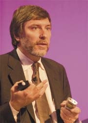
Thus, Dr Jean-Luc Vanderheyden underlined the potential of molecular imaging, during an interview: Meike Lerner, European Hospital. ‘If factors that cause cardiovascular diseases could be made to ‘glow’, he explained, ‘then anomalies could be detected before an event occurs.
Currently we are working on the ability to detect patients who would be asymptomatic,’ Dr Vanderheyden explained. ‘There are a couple of cardiology programmes; one, for example, involves a tracer – a molecule – to image the adrenergic receptor – to which norepinephrin binds. Making an imbalance in these receptors visible would enable us to identify the risk of congestive heart failure. The clinical studies are very promising and we are confident that, in the near future, we will be able to identify those millions of people with a silent heart disease who might be at risk of a congestive heart failure.
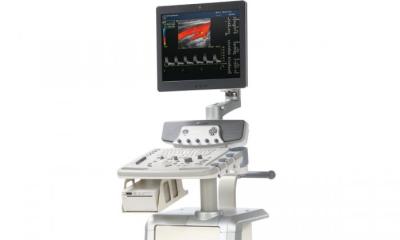
On the other hand, we are working on progressing imaging technologies, in particular the special contrast agents that correspond with the tracers. With VCT (very rapid CT) we can image the heart in five heartbeats and ten seconds with an extremely low dose of radiation (70% reduction compared with the normal dose). The combination of special contrast agents and advanced technology makes it possible to look particularly at stenosis in a cardiac blood vessel, and therefore identify an at risk patient.’
Do some imaging technologies look more promising than others for molecular imaging in cardiology?
‘In this field we are not concentrating on any particular modality. It is more important to understand the disease, and then look for the imaging modality that will provide the best answers. In oncology we work a lot with PET. GE also has a pilot project for prostate cancer with MRI. In cardiology most of the work has been with SPECT and SPECT tracer, which show perfusion and the patients at risk of heart attacks. But these technologies alone can only show risk in patients already identified due to an event or chest pain. Only if we add this special molecule as a tracer can we see anomalies in advance. To come to the point: talking about molecular imaging really means the combination
of a tracer – a molecule, and sophisticated equipment that can detect the signal of the molecule. Of course, software solutions must be developed that allow greater ease of visualisation and help doctors in their diagnoses. Our objective is to use all possible modalities to identify at risk patients. It is less critical which modality is used, from VCT, ultrasound or in-vitro diagnostics.’
Such sophisticated technologies and advanced software always need well-trained doctors to handle those technologies and make sound diagnoses.
‘Yes, education is vital. That’s why we observe all and everything during clinical trials, and then record this information connected with the procedure in a manual – for example, patient preparation. It’s not just doctors who need to rethink, with molecular imaging patients will also play a greater role and have greater responsibility. Last, but not least, healthcare systems must shift from treatment to prevention and recognise that it will be more important to deal with patients when they do not have heart attack or congestive heart failure symptoms, rather than waiting for symptoms to arrive, then sending them to an emergency room to decide the best treatment.
When we talk about molecular imaging in cardiology we are really talking about a paradigm shift from treating symptoms to preventing disease. Looking at the state of play today, I think we can expect a lot from this field. Already, sophisticated techniques have improved to a lot – two or three years ago we couldn’t see the things we see today. So, there is continuous progress and if we look further into the future we should be able to document metabolic changes that would be negative for ischaemia, including perhaps the effects of hibernation – and certainly we should be able to demonstrate that alteration of the sympathetic nerve function, or the adrenergic ones that result in coronary diseases.
And maybe – in about five years of using molecular imaging – we’ll be able to look at plaque characterisation, including the identification of the vulnerability of that plaque or additional receptor characterisation that can lead to drug therapy. And the last would be biomarkers, to perhaps identify some of the recorded disturbance instability that could be associated with atrial or ventricular arrhythmia.
So, quite a lot of things will be in the works that wouldn’t have been possible without the combination mentioned, as well as the sophisticated technologies and suitable target agents.’
30.05.2007