DISCHARGE
Less unnecessary cardiac catheterisations in the future
Cardiac catheterisation is the gold standard for diagnosing coronary artery disease (CAD), the main cause of death worldwide. More than 3.5 million invasive coronary angiographies (ICAs) are performed in the European Union each year, tendency rising. Nearly 60 percent of these minimal invasive examinations do not result in further treatment, since the patients do not have obstructive epicardial coronary artery stenosis. For these patients non-invasive cardiac computed tomography (CT) could be advantageous. The randomised study DISCHARGE currently examines for which patients with suspected coronary artery disease based on stable chest pain, CT or ICA is preferable. Marc Dewey, Heisenberg Professor of Radiology at the Charité University Hospital in Berlin, coordinates the study, taking place at 25 clinical sites in 16 European countries.
Aiming for novel guidelines
More than 3500 patients will be recruited within the next two years for the study from now on, with a maximum follow-up of four years. The major goal of the trial is to determine whether CT helps to reduce myocardial infarction, stroke, and cardiovascular death. Procedural complications will be a secondary outcome. The clinical trial is part of the multinational research project DISCHARGE, which has been granted six million euros through the 7th Framework Programme of the European Union (EC-GA 603266). The DISCHARGE consortium consists of 30 members in 18 European states. “Ultimately, DISCHARGE aims to provide the basis for new guidelines in cardiac imaging,” says Prof. Dewey. “Therefore, we are closely collaborating not only with clinical sites, but also with non-clinical partners to optimise the impact of the study for the benefit of the different European health systems.”
CT may improve outcomes
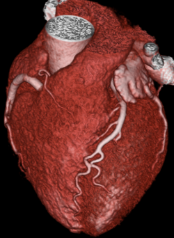
ICA allows minimally invasive treatment of coronary stenosis during the examination. But the invasive technique bears a risk of serious side effects for about 1 percent of the patients. “For patients with suspected CAD with a low-to-moderate risk (10%-60%), non-invasive CT might provide a better risk/benefit ratio in favour of the patient,” says Prof. Dewey. “Early detection and improved characterisation of coronary plaques in the entire coronary artery tree is also very well possible with CT. Certain unique high-risk plaque features have been also been shown to predict subsequent events and outcomes if assessed by CT.” It is however unknown, whether such high-risk plaques should lead to recommend intensified risk factor modification or certain medications to improve outcomes.
Additional diagnostic benefit
CT imaging also allows the examination of tissues surrounding the heart: lungs, oesophagus and spine may be checked for instance. This may result in a diagnosis explaining chest pain and suggesting appropriate treatment, which could be overlooked by ICA. “ICA is the best way to treat known CAD. But in a situation where ruling out diagnosis of CAD is likely, CT, with its tremendously improved image quality, might prove to be the best method available,” says Prof. Dewey. Currently, CT has little role in diagnosis of CAD and is not reimbursed for this purpose in most European countries yet. There are other imaging modalities to rule out CAD besides CT such as stress MRI, PET/CT, SPECT and stress echocardiography. These perfusion-imaging tests enable to search for stress-induced ischemic myocardial areas and play an important role in clinical decision-making in the DISCHARGE trial in case of anatomic coronary stenosis found by CT with unclear functional relevance.
Outlook on the future of cardiac diagnostics
Dedicated research groups in Europe are working on all of these non-invasive cardiac imaging techniques, to further improve these diagnostic tests from a technical and clinical perspective. “The main goal would be to develop a comprehensive imaging test that allows accurate stenosis detection, characterisation of coronary plaques and myocardial perfusion quantification,” says Prof. Dewey. “CT with its wide availability and high diagnostic accuracy and more costly hybrid imaging techniques such as PET/CT, are most promising to comprehensively assess CAD.”
Source: ESR
16.10.2015