Looking beyond the lungs: don’t forget the cardiac structures!
So close yet so far away! A fitting description of many diagnostic examinations of heart and lungs. Modern imaging modalities allow these two neighbouring organs to be evaluated together, nevertheless it is rarely done.
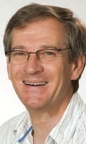
A very outdated approach as Professor Dr Karl-Friedrich Kreitner, Consultant at the Clinic and Policlinic for Diagnostic and Interventional Radiology at the University Hospital Mainz, explains: “Many diseases that start in one system sooner or later affect neighbouring organs. Therefore we have to look at heart and lungs as a functional unit that needs to be assessed accordingly.”
This modern approach is made possible by the technological advances in computed tomography during the past years. “Today we can perform a chest scan with prospective ECG triggering without having to increase radiation exposure. This allows us to visualize the lung and cardiac structures in sharp detail”, says Professor Kreitner. An integrated examination is particularly valuable when lung diseases can affect the heart, for example pulmonary hypertension. But there are also many cardiac diseases that impair lung function such as comple heart defects or conditions that limit cardiac functions such as cardiac insufficiency or hypertension. The close look at heart and lungs alike has many advantages, Professor Kreitner underlines:
“Take a smoker with COPD: with a prospectively triggered chest scan we can also evaluatethe coronary arteries for calcifications, stenoses or even infarctions.” This new approach however has not yet taken hold in clinical practice. Very few facilities have a so-called cardiothoracic imaging center where the findings of different disciplines are discussed. “Specializations within radiology,” Professor Kreitner points out, “continue to be highly inflexible. Moreover, the cardiologists frequently consider diagnostics of the heart their proper domain. This does not make sense at all and I expect things to change substantially in the short run. Organizations such as ESTI have already picked up on the trend and have integrated cardiac diagnostics in their scientific programme, an important step which might lead to ESTI and the European Society of Cardiac Radiology offering a joint congress.”
Obviously additional information requires additional know-how that the radiologists need to acquire. But Professor Kreitner urges his colleagues to take on the challenge; indeed he is convinced that the integrated evaluation is a great opportunity for radiology to showcase its core competencies: “With this additional knowledge we could show that cardiac imaging is the task of radiologists rather than cardiologists. Cardiothoracic imaging offers us the opportunity to clearly reject the image of mere ‘button pushers’ that has been haunting us. The technological developments force us to take a competent stand on the issue.”
Next to CT it is MRI that is on its way to conquer thoracic imaging. It remains to be seen however to what extent MRI will be able to offer meaningful visualizations of the lungs. But there are interesting developments. “The clinical value of several functional approaches, for example with regard to pulmonary haemodynamics, perfusion orventilation, needs to be discussed. Today thoracic MRI is still rather involved, nevertheless I do expect larger studies soon that will urge us to think about diagnostic algorithms,” the radiologist predicts.
Until then, Professor Kreitner recommends, his radiology colleagues ought to exploit the potential of CT and look beyond the lungs: “Take a look at the mediastinum and the cardiac structures. Don´t forget them!”
####
Profile
Since 2008, Dr Karl-Friedrich Kreitner is Professor of Radiology at the Departmentof Diagnostic and Interventional Radiology of Johannes-Gutenberg University in Mainz, Germany, where he has been teaching imaging since 2004.
To date Kreitner, who has two children, has published more than 180 articles in peer reviewed journals and 22 contributions to books. He is co-author of the book Cardiac Imaging: A Multimodality Approach (Thieme Publishers, New York, 2008). Professor Kreitner is a member of several professional associations, inter alia RSNA, Deutschen Gesellschaft für Kardiologie-Herz- und Kreislaufforschung, PROFILE ESR and ESTI.
19.06.2012