Sponsored • Testing Tissue
Cone Beam CT for the breast
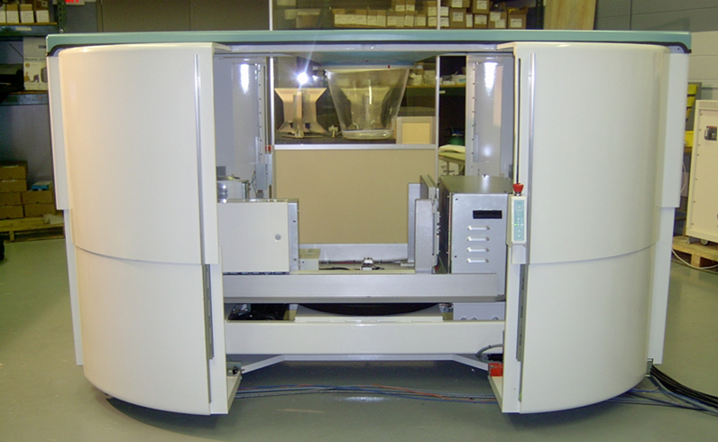
The technology can acquire hundreds of images in only ten seconds, producing true 3D images to enable a fast procedure with excellent patient comfort, the manufacturer explains.‘Optional accessories for KBCT include a biopsy bracket to enable KBCT-guided biopsies of suspicious lesions, and a collimator, used to limit the X-ray beam to the area of interest. The biopsy bracket provides 3D targeting at comparable or lower radiation exposure compared to stereotactic guided biopsy.’
A view like no other

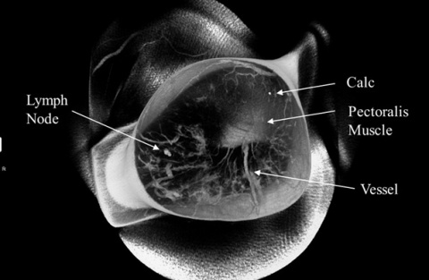
The breast CT images have less distortion than mammography and the system is optimised to differentiate between the breast’s soft tissue and cancer tissue, Koning points out. ‘These images will be very different from 2D mammograms. They’re truly 3D images of the entire breast from any orientation. You can scroll through the slices (up and down, left and right) and get a unique view of the breast like never before. ‘It gives doctors tremendous freedom in how they look at the interior of the breast and evaluate its structures. It’s almost like seeing the anatomy itself.’
No breast compression
As Ruola Ning PhD, Koning’s President and Founder, a pioneer and leading expert in Cone Beam CT Technology and sole inventor of cone beam breast CT technology, confirms: ‘KBCT represents a revolutionary advancement in breast cancer diagnosis.’This is the first commercially available 3D breast CT scanner designed specifically to image the entire breast with a single scan, without compression of the breast tissue – which means this procedure is far more comfortable for patients than regular mammography. Additionally, Koning adds that there is less radiation exposure than during a CT exam of the entire chest, because here only the breast is exposed to X-rays.
06.03.2015


