Article • 100th birthday of Sir Godfrey Newbold Hounsfield
The legacy of the man who pioneered computed tomography
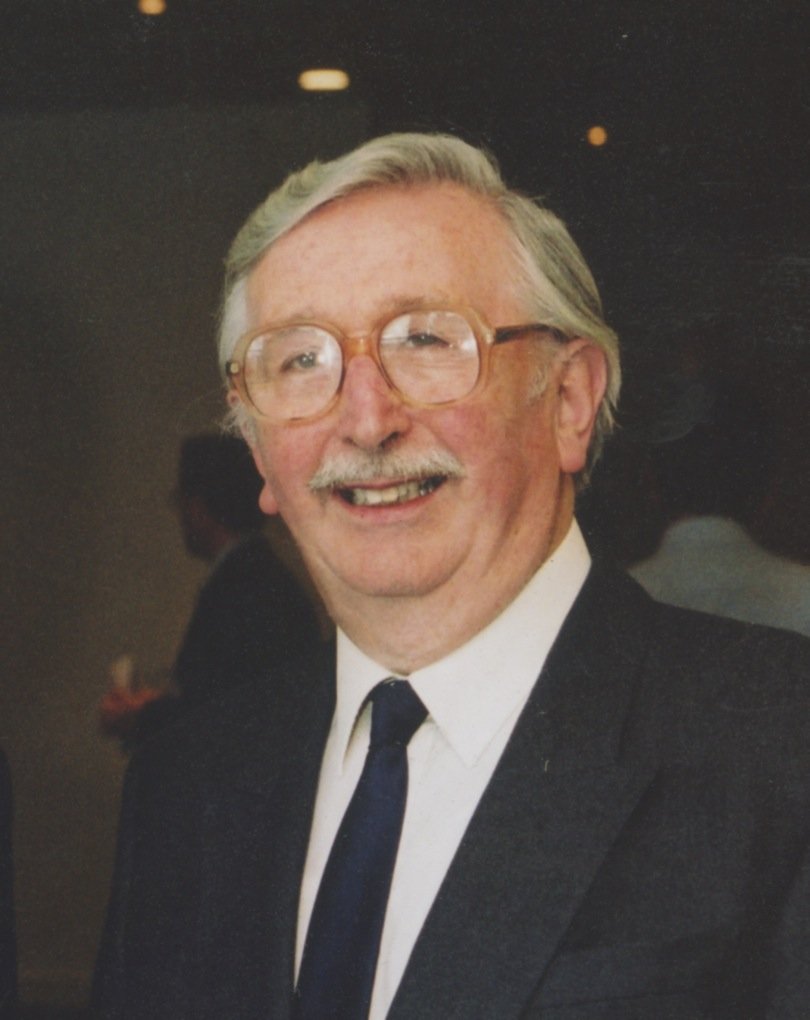
On the centenary of his birth, Mark Nicholls reflects on the life and legacy of Nobel laureate Sir Godfrey Newbold Hounsfield, the man who pioneered computed tomography.
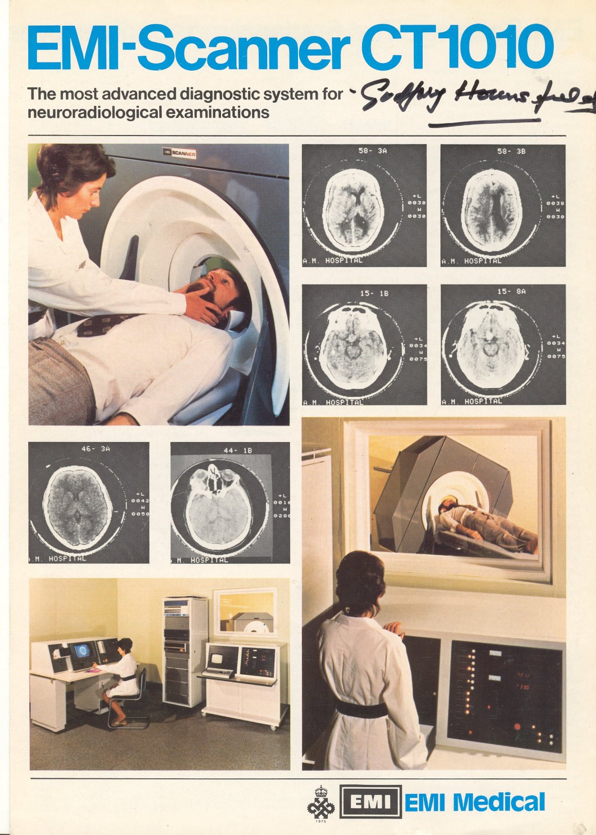
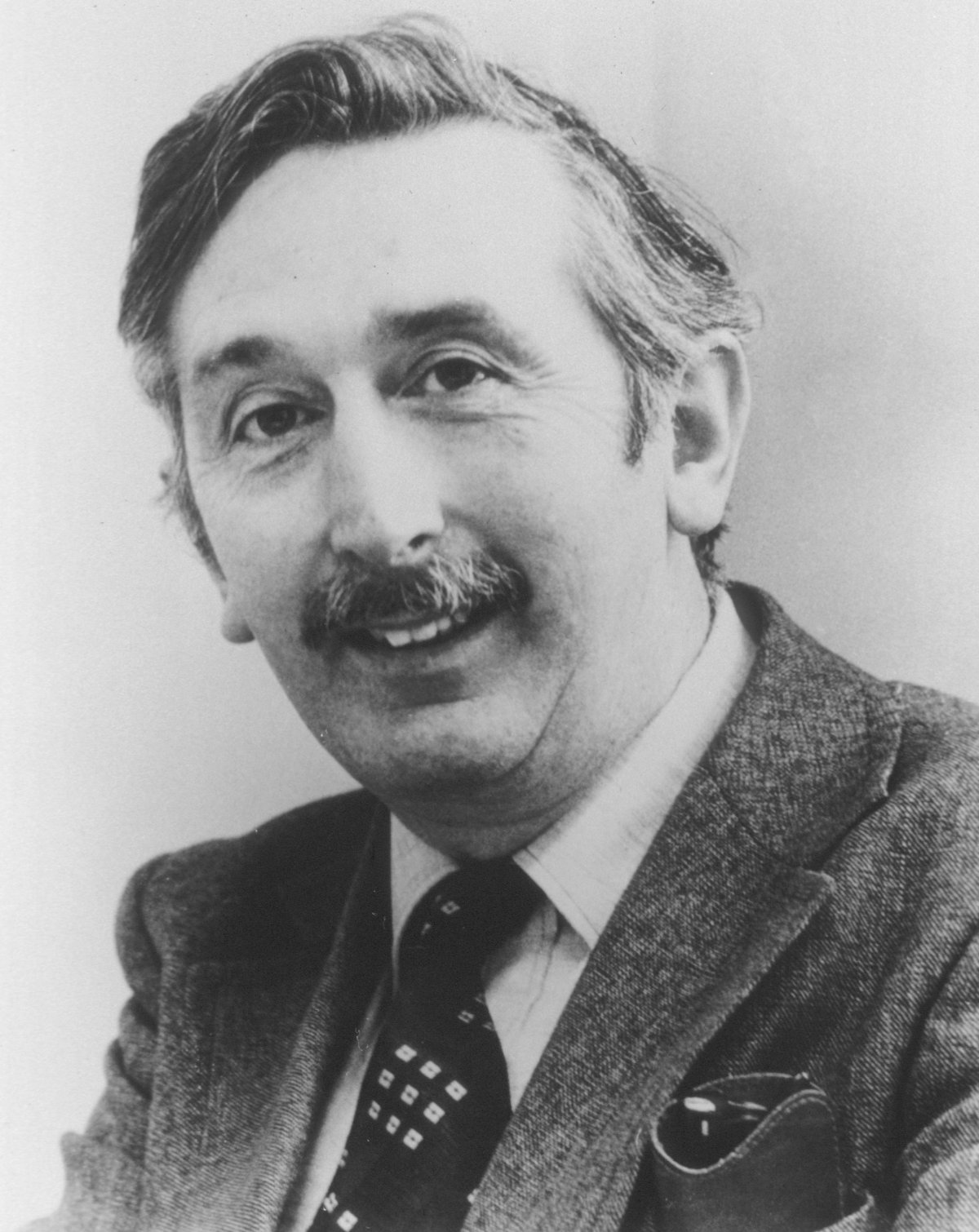
Image kindly provided by Prof Adrian Thomas
It was a discovery that came from a moment of inspiration during a country walking holiday; the idea that one could determine what was inside a box by taking X-ray readings at all angles around the object. From that, Sir Godfrey Newbold Hounsfield set to work constructing a computer that could take input from X-rays at various angles to create an image of the object in “slices.” The concept of CT (computed tomography) – and a technological breakthrough that would revolutionise medical diagnostics from the outset – was born.
This year sees the 100th anniversary of Sir Godfrey’s birth on August 28, 1919, and also marks the 40th anniversary of the award of the Nobel Prize in Physiology or Medicine – jointly with Allan M. Cormack – for the “development of computer assisted tomography.”
Today, it is widely acknowledged that the work of Hounsfield and Cormack has become a “foundation” of how patients are managed and assessed in all specialities with CT imaging instrumental in the diagnosis of cardiovascular disease, acute neurological, chest and abdominal conditions, as well as trauma.
The introduction of modern imaging
Reflecting on his legacy, Professor Adrian Thomas described Sir Godfrey’s contribution to modern day medical assessment as “remarkable.”
“At a stroke, investigative medicine was transformed,” he said. “The approach was entirely different to that being pursued by the X-ray industry of the period. The scanner transformed medical investigations, and transformed treatments – both image-guided interventions and radiation therapy.” Professor Thomas, a radiologist in private practice and visiting professor at Canterbury Christ Church University teaching postgraduate radiography, is also co-author of the 2012 biography “Godfrey Hounsfield: Intuitive Genius of CT” with Liz Beckmann, Richard Waltham, who was an engineer on Hounsfield’s team, and Stephen Bates, described as Godfrey’s ‘right-hand man.’ Basically, says Professor Thomas, Hounsfield’s legacy is “the introduction of modern imaging.”
“Whilst there is no monument to Hounsfield, it could be said that his monument is the presence of a CT scanner (and often several) in every A&E/Emergency Department.”
Professor Thomas described Sir Godfrey as someone who had “an enquiring mind, was hard working, and a great leader and motivator of his team” and was acutely aware of the impact of his discovery and what it would lead to.
“He was absolutely charming, and was very humbled and pleased to receive the many letters that patients who had benefitted from his invention,” he added.
Nobel Prize
Within 21st century medicine, CT is a leading hi-tech diagnostic and evaluation tool, having evolved from the development of computer assisted tomography in the 1970s, for which Godfrey Newbold Hounsfield and Allan M. Cormack (1924-1998) – working independently – were awarded the Nobel Prize in Physiology or Medicine for 1979.
With CT, the X-ray film is replaced by sensitive crystal detectors, and the signals emitted by amplifiers when the detectors are struck by rays are stored and analysed mathematically in a computer. The computer is programmed to reconstruct an image of the examined cross-section by solving a large number of equations. With high sensitivity, CT can detect very subtle variations of tissue density, meaning that the density resolution is exceptionally high. The process causes little discomfort to the patient, though there is an element of radiation exposure and intravenous injection of radio contrast.
Sir Godfrey was raised on a farm in Nottinghamshire, England, the youngest of five children. He joined the RAF at the start of World War II as a Radar Mechanic Instructor and after the war attended Faraday House Electrical Engineering College in London before joining EMI, working on radar and guided weapons.
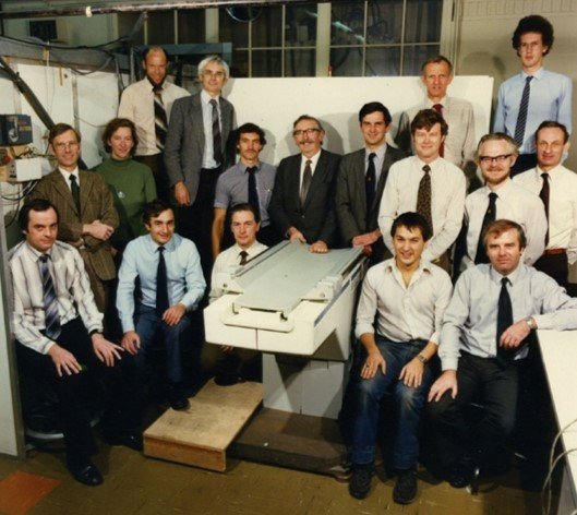
He became particularly interested in computers, which were then in their infancy, and in 1958 led a design team building the first all-transistor computer to be constructed in Britain, the EMIDEC 1100.
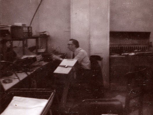
Later, transferring to EMI Central Research Laboratories, the idea occurred to him in 1967 which was eventually to become the EMI-Scanner and the technique of computed tomography.
Pattern recognition
Mrs Beckmann, who joined EMI in January 1977 as part of the technical sales support team, explained why the first CT examined the skull and diseases of the brain.
“When Godfrey started, he was not looking for a medical image, it was about pattern recognition and he wondered if he could recognise the contents in a box by taking a large number of readings through the box, but then while on a holiday he was talking to a doctor who said the ‘box’ they were having trouble looking inside was the brain so adapted the original concept to looking at the brain.”
Later, she got to know him better as President of the British Institute of Radiology (BIR) when the organisation began staging annual Godfrey Hounsfield lectures.
She is in no doubt as to the impact of his contribution: “His was one of the greatest, if not the major, contribution to imaging - if not in the whole field of medicine - in the 20th century. We would not have areas of diagnostic, surgical, or therapeutic medicine without CT. Everything from computer aided design to artificial intelligence used in medicine today, has links to this early application of computers to the imaging world.
“Because of his major contribution, we should recognise the centenary of his birth in the way we would with any great people – he was not a brilliant professor of engineering, but he was intuitive. He was certainly unique and he did not conform to norms but the Nobel prize was well deserved.”
When the Nobel Prize was awarded to Hounsfield and Cormack, their discovery was heralded as a “revolutionary radiological method, particularly for the investigation of diseases of the nervous system.”
Cormack, who was professor and head of the institution of physics at Tufts University in Medford, Massachusetts, USA, was the first to analyse the conditions for demonstrating a correct radiographic cross-section in a biological system, publishing his analysis in 1963.
Hounsfield, however, as chief of the medical research division of Electric and Musical Industries (EMI) in Middlesex, England, is regarded as the central figure in invention of computer-assisted tomography in constructing the first practicable CT system for medical care. Unaware of Cormack’s contributions, he developed his own method for reconstruction of the image.
A revolutionary diagnostic instrument
When introduced into medical care, the impact of CT as a revolutionary diagnostic instrument was recognised from the outset.
The Nobel citation of 1979 for Sir Godfrey read: “…he obtained results which in one blow surprised the medical world. It can be no exaggeration to maintain that no other method within X-ray diagnostics has, during such a short period of time, led to such remarkable advances, with regard to research and number of applications, as computer-assisted tomography.”
Forty years on, Professor Thomas says that statement remains as true in the 21st century as it did then.
“Modern doctors cannot now imagine how medicine was practised prior to the introduction of the EMI CT Scanner,” he said. “Prior to CT, radiologists saw things by displacement and in an indirect way; before CT, we had invasive diagnosis and invasive treatment. Now, the modern paradigm is non-invasive diagnosis on the basis that you have minimally-invasive therapy, but you can only have minimally-invasive therapy if you have accurate diagnosis. Clinically, CT completely transformed the way doctors manage patients.”
CT continues to evolve today: the modality is fast, now uses lower doses of contrast, can show rapidly moving structures (such a coronary arteries), has a role in guiding biopsies and therapeutic interventions and is being further developed via artificial intelligence.
Recommended article
40 years of CT scanning
Forty years ago an article was published that would change medical practice. In the British Journal of Radiology, English electrical engineer Godfrey N Hounsfield described how he had made a patient’s brain visible non-invasively by evaluating a large number of X-ray images of the skull taken from different directions.
Team player and loner
EMI had the Beatles at the time and one of the great myths is that the money made by the Beatles helped fund the CT scanner – that is not the case.
Adrian Thomas
Mrs Beckmann, currently chairman of the British Society for the History of Radiology (BSHR), said that many of the modern forms of CT can be traced directly back to Sir Godfrey’s early ideas.
“In the last few years CT has started to use intuitive reconstruction to create images, but it is worth reminding people that those were the methods that Godfrey Hounsfield and his team initially used but the algorithms were not smart enough and the computers not fast enough back then. Spiral CT can also be traced back to Godfrey; the drawings are there in his original notebooks.”
She said he did recognise the impact his work had but she added: “He was very modest, he did not feel he deserved the accolades or enjoy the publicity, he was much happier siting in the corner of his office, but at the same time he was a team player and he did enjoy socialising with his colleagues. Once, when we took him to the BIR lecture in is name in Manchester, he was booked into a four-star hotel, but he insisted on staying with everybody else in a cheaper hotel and with people he knew – that was very much Godfrey Hounsfield.”
There are, however, several myths surrounding the development of the EMI scanner.
Professor Thomas said: “EMI had the Beatles at the time and one of the great myths is that the money made by the Beatles helped fund the CT scanner – that is not the case. In terms of funding it, there was no huge budget for the EMI scanner, it was done on an absolute shoestring.”
The Sir Godfrey Hounsfield lecture
The centenary of Hounsfield’s birth will be commemorated by the Sir Godfrey Hounsfield lecture during the BIR’s annual congress in November and be delivered by Professor Andrew Scarsbrook, Consultant Radiologist and Nuclear Medicine Physician, Leeds Teaching Hospitals NHS Trust, on the subject of “PET/CT: From evolution to revolution.”
CT scanning was introduced into medical practice on October 1, 1971, with a successful scan on a cerebral cyst patient at Atkinson Morley Hospital in Wimbledon, London. From there, Sir Godfrey continued to improve CT scanning, introducing a whole-body scanner in 1975.
A Fellow of the Royal Society and knighted in 1981, he retired in 1984 and died, aged 84, on August 12, 2004, with his name immortalised in the Hounsfield scale, a quantitative measure of radiodensity used in evaluating CT scans.(mn)
28.08.2019