"Children are at the other end of physics"
Small patients challenge today's imaging
As in every medical field, children have special needs in radiology. Increasingly aware of this -
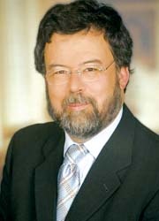
and just as they must adapt scanners for increasing numbers of obese patients - manufacturers are sharing lively exchanges with practitioners to develop the most advanced equipment available for very small patients. At the Medical University in Graz, Austria, which has taken a lead in this endeavour, Meike Lerner asked Professor Erich Sorantin (below), of the Clinical Department for Paediatric Radiology, about the challenges in this field and ways to overcome them.
‘The main difference is definitely the size: With children we are, in the most extreme cases, talking about a body weight of 400 grams compared with, for example 120 kg in a grown man – this corresponds to a mass factor of 300. Additionally, in paediatric radiology we are familiar with a whole range of indications and cover all areas: orthopaedics, cardiology, oncology but also neurology. We really do see the patient as a whole.
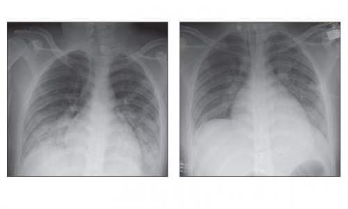
‘Up to just a few years ago, imaging in paediatrics was tagged on to adult imaging. For a long time we tried to adapt the possibilities in adult diagnostics to children. However, although there is a common technological development, with children we have to work with very different factors and base our work on very different parameters. For example, for a simple lung X-ray we had to adapt the software for the post-processing of an examination especially to work with children, because we couldn’t work with the standard solution. We then developed a solution with the manufacturer. It took us 18 months to adapt the first digital radiography system to the needs of children in 1995. With a further installation in 2003, a flatbed scanner for projection radiography, we also had to do a lot of work to adapt the post-processing software. The company – Siemens at the time – then introduced their own release to the market.
‘We should look at it this way: Children are at the other end of physics. Everything we do here will ultimately benefit adults. And the new developments, such as PET/CT or hybrid systems respectively, generally have to be evaluated in a very different way for children. For example, with children we have to work with low contrast resolutions because of the lower proportion of fat. In short, we have to put the cart before the horse and start with the smallest – they need the largest equipment.’
What equipment is suitable for very small patients?
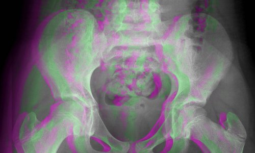
‘With X-ray systems there is a relatively simple model: We know that you can “simulate” the abdominal mass of children fairly well using Plexiglas and we also know how to measure the relevant diameters subject to age. By choosing the appropriate Plexiglas pane (available in panes of 1cm) we can take phantom images for different ages and parameter tests. Then, for example, we fit a stomach tube, endotracheal tube or a strip of gauze to see whether you can actually see these on the X-ray image. This means that, with the help of a simple phantom, we can determine whether the equipment is suitable for children.
‘Of course, we also work with more specific procedures, such as the Wellhöfer phantom to carry out differences in the thickness or quantifications, or to vary the dose, and to analyse dependent parameters, such as image noise. In Graz we’ve already developed a programme specifically for these tests, which we can use to carry out these adjustments.
‘In addition, we have to consider physiological changes: a child triples its weight in its first year of life but only doubles its height. Further changes are that the bones calcify based on age – younger children have lower bone density and a higher proportion of cartilage etc. These changes must all be fed into the examination protocols. Therefore we require data from very different medical disciplines to bring them together. From this process the result is certain protocols, which we try to standardise. The CT scans in preparation for funnel chest surgery, for example, in 99% of cases are carried out in the ultra-low dose mode because this is often only about the deformity.
‘Thanks to imaging we can carry out many more examinations these days, but now these exams must become even more subtle because we need more precise therapy gradings
‘It is decisive that developments – also those in the industry – lead to a point where children are no longer seen as small adults but where specific solutions are developed for this group of patients.’
01.05.2009