Image source: ARRS
News • DSCT benefits
Coronary CTA for acute chest pain emergencies: Single- vs. dual-source scanners
According to an accepted manuscript published in the American Journal of Roentgenology (AJR), using a dual-source CT (DSCT) scanner for coronary CTA can facilitate clinical processes by eliminating the need to administer beta-blockers for heart rate control while decreasing nondiagnostic examinations.
The findings indicate the potential role of a DSCT scanner with high temporal resolution to help expedite clinical processes in the emergency department (ED) setting
Young Joo Suh
“The findings indicate the potential role of a DSCT scanner with high temporal resolution to help expedite clinical processes in the emergency department (ED) setting,” concluded corresponding author Young Joo Suh, MD, PhD, from Korea’s Research Institute of Radiological Science at the Center for Clinical Imaging Data in Seoul.
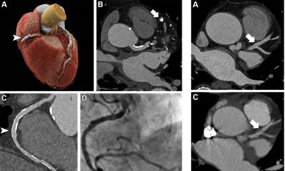
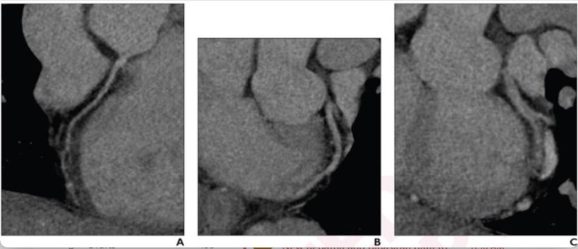
This AJR accepted manuscript included 509 patients (mean age, 52.1 years; 283 men, 226 women) at low-to-intermediate risk for acute coronary syndrome who underwent coronary CTA for acute chest pain in a single ED during the off hours of March 1, 2020 to April 25, 2022. A total 205 patients initially underwent CTA using a 64-detector single-source CT (SSCT) with heart rate control—oral beta-blocker administration if heart rate was faster than 65 beats per minute. Following scanner replacement on April 26, 2021, 304 patients underwent CTA using a third-generation DSCT without heart rate control. Cohorts were compared via length of stay (LOS) in the ED, as well as CT completion time (i.e., from ordering CTA to complete acquisition). Additional endpoints for Suh et al. included image quality and nondiagnostic examinations based on radiology reports.
Ultimately, DSCT without heart rate control—compared to SSCT with heart rate control—showed no difference in ED LOS (505 vs. 457 minutes; p=.37), but shorter CT completion time (95 vs. 117 minutes; p<.001), lower frequency of nondiagnostic examinations (1.6% vs. 6.3%, p=.01), and no difference in cardiology consultation, angiography, disposition, or revascularization (p>.05).
Noting an array of factors, especially patient disposition and onboarding processes, can affect overall length of stay, “the reduction in CT completion time through use of the DSCT system can still provide overall benefit in the ED setting,” the authors of this AJR accepted manuscript added.
Source: American Roentgen Ray Society
03.03.2023