Three questions for WFUMB Honorary Lecturers …
World of Ultrasound met Prof. Ioan Sporea, Prof. Gebhard Mathis and Prof. Byung Ihn Choi to ask them three questions.
Can liver elastography replace liver biopsy – today or sometime?
On 27 August, University Professor Ioan Sporea MD, Head of the Department of Gastroenterology and Hepatology at the University of Medicine and Pharmacy in Timisoara, Rumania, will deliver the EUROSON lecture: Can liver elastography replace liver biopsy – today or sometime?, which particularly focuses on Acoustic Radiation Force Impulse (ARFI).
1. What exactly is ARFI and how does it work?
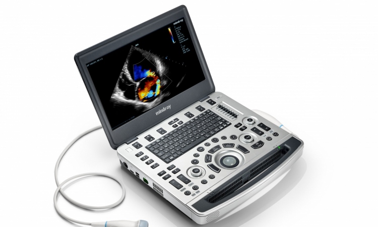
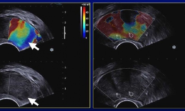
Prof. Sporea: ‘ARFI is a new elastographic method that assesses tissue stiffness. The ultrasound probe automatically produces an acoustic “push” pulse that generates shear-waves, which propagate into the liver. Using image-based localisation and a proprietary implementation of ARFI technology, shear wave speed may be quantified, in a precise anatomical region, focused on a region of interest, with a predefined size, provided by the system. The velocity is proportional to the tissue stiffness, with faster wave progression occurring through stiffer material and is measured in meters/second.’
2. Compared with other imaging methods, what are the advantages and disadvantages of ARFI?
‘The ARFI “real-time” evaluation method can be used in patients in whom liver stiffness measurements by Transient Elastography could not be obtained (because the ARFI location measurement can be chosen under direct ultrasound guidance), and also in patients with ascites.
‘ARFI is also a rapid method to assess liver fibrosis, totally free of adverse events, comfortable for the patient and for the examiner (with a mean duration of approximately five minutes).
‘Another advantage is that the LS measurements can be made with a device integrated into an ultrasound machine (already existing in some ultrasound laboratories). So, ARFI measurements can be obtained immediately after an ultrasound liver evaluation and information regarding the severity of liver fibrosis is available on the spot, without having to buy another machine.’
3. Are there recent developments or innovations in this elastography method?
‘There are several published studies that proved its value for fibrosis assessment in chronic hepatitis and cirrhosis. Also, our group has published a study regarding ARFI elastography for the differential diagnosis of normal and diffuse thyroid pathology.
‘Ongoing studies are also evaluating ARFI for the assessment of diffuse thyroid pathology of hepatic and thyroid nodules.’
Ioan Sporea
Born in Romania in 1956, Professor Ioan Sporea MD PhD graduated in 1981 at the University of Medicine and Pharmacy of Timioara. He specialised in internal medicine and gastroenterology, with an emphasis in non-invasive staging of diffuse liver disease, multimodal treatment of hepatocarcinoma, ultrasound of the digestive tract etc., and now heads the Department of Gastroenterology and Hepatology at the same university.
He is President of the Romanian Society of Ultrasound and Elected-President of the Romanian Society of Gastroenterology and Hepatology.Prof. Sporea is also author and co-author of 118 original papers and more than 700 published studies.
Lung-US: A newly arriving technique
In his WFUMB Honorary Lecture, Professor Mathis will focus on Lung-US: A newly arriving technique.
Ultraound is indeed developing into an indispensable modality, for example to diagnose pneumothorax, infections or embolisms, and shows a sensitivity comparable to spiral CT. In the WFUMB publication ‘World of Ultrasound’ Prof. Mathis answered the three of the most important questions on this subject.
Which indications is ultrasound suitable for?
Prof. Mathis: ‘There was an international consensus conference on the subject last year where the indications – based on current experience – were clearly defined: Its use in cases of suspected pleural effusion has long been known. However, the use of ultrasound to confirm or eliminate pneumothorax is less known and common, although very effective.
‘It can also be used to detect pneumonia, with a sensitivity of 94%, although it cannot be used to rule out pneumonia. Pulmonary embolism also becomes clearly visible through ultrasound in many cases, with a sensitivity of 80%. Only central pulmonary embolisms remain hidden. By combining procedures, such as echocardiography and thoracic ultrasound in the case of compression ultrasonography of the leg veins, we also achieve a sensitivity of 92%. Pulmonary embolism cannot be ruled out, although this is not possible with any other imaging procedures either, including spiral CT.
‘Generally, ultrasound allows us to show consolidations under the pleura, no matter whether these are infectious, embolic or benign processes. In the case of tumours for instance ultrasound guided aspiration is extremely accurate. The recommendation in the consensus paper states that in certain clinical situations, as described above, or where CT is counter indicated – for example in the case of kidney insufficiency, contrast medium allergy or pregnancy – ultrasound should be used first.’
Which technological developments have a positive impact on lung diagnosis via ultrasound?
‘Contrast medium ultrasound, increasingly being used, is of particular interest. As infectious consolidations strongly concentrates, small, subpleural lesions can be well and safely distinguished from embolic changes. Such diagnosis is not at all possible with CT; ultrasound has the clear advantage here.
‘The new mobile, handy devices have also opened up new areas of application. There is a real change of paradigm here, because the probe can be used almost like a stethoscope all the time. In the US, ultrasound is therefore increasingly becoming part of the standard equipment for emergency doctors, who have rediscovered this procedure for themselves – a trend that’s also becoming apparent in Europe. I’m convinced that a lot will happen in this field in the coming years.’
Are there limitations to this procedure?
‘The biggest limitation is air, which takes away the image. Otherwise, abdominal, cardiac and vascular ultrasound diagnosis requires a certain amount of experience to achieve the right result. We recommend a course that teaches the theoretical basics. Other than that I actually see more opportunities than limitations for lung diagnosis via ultrasound.
Gebardt Mathis
Following employment as an internist at the Hohenems Hospital, the Medical Clinic St. Gallen and the Vienna University Hospital, and an eight-year professorship at the Innsbruck Medical University, since 2006 Professor Gebhard Mathis has run his private practice in Rankweil, Austria.
During his career, the 61-year-old has been the recipient of the Durig Böhler Prize four times. He has published 130 scientific contributions and authored various specialist books focusing on, for example, lung, pleura, chest and gastrointestinal ultrasound.
The imaging of liver cancer: recent advances
What importance do global cooperation and, especially the gathering of experts at WFUMB hold for you?
Prof. Choi: ‘WFUMB is the largest and prestigious organisation consisting of six regional federations of ultrasound (US) with more than 55,000 members, and has a Congress of WFUMB which should be a representative meeting of ultrasound, not only in quantity but in quality. This meeting should be an ideal place to gain recent knowledge and cutting-edge information of US and to meet old and new friends in the field of US and radiology as well.
‘Therefore, each regional federation has to cooperate with world federation to promote this meeting and encourage US specialists, particularly young people, to participate in this meeting. Fortunately, the meeting will be held in Vienna in nice season. Vienna is a very rich cultural city in music, fine art, architecture, and history. So, I expect that many people will attend and enjoy this scientific meeting as well as … Vienna.’
Will contrast agents be deployed in liver imaging?
‘Recently, for liver imaging diagnosis, less invasive imaging examinations, such as US, CT and MR, have developed dramatically. The recent development of CT includes multidetector-row CT (MDCT) and dual energy CT. With the new CT machine, CT imaging has developed from 2-D to 3-D imaging, and the recent addition of time axis to the 3-D imaging, so called 4-D imaging, has become available Moreover, research is developing in functional imaging, such as tissue blood flow analysis by perfusion CT. The recent advance of MR imaging is in fast MR imaging technique, high Tesla MR, 3-D gradient echo sequence for T-1 and tissue specific MR contrast media, especially hepatic tissue specific contrast medium.
‘Recently developed ultrasound technologies include four dimensional US, elastography, and contrast-enhanced US with various microbubbles. CEUS has been used in various clinical indications of liver disease. CEUS is now recognised as a useful imaging modality for non-invasive diagnosis of small newly detected liver nodules during hepatocellular carcinoma (HCC) surveillance, and provides an accurate differentiation between benign and malignant liver tumours, which is critical for the adequate management of these patients.
‘CEUS is also useful for guidance and follow-up for loco-regional therapy of HCC. In addition, the recently introduced new contrast agent Sonazoid, which is phagocytised by Kupffer cells, is a dual function agent not only for vascular phase but for post-vascular phase (Kupffer phase). So, Sonazoid-enhanced US can improve the detection and characterisation of liver tumours, thus improving the management of liver cancers.
New procedures such as Selective Internal Radio Therapy (SIRT), thermo ablation or chemo embolisation play an increasing role in hepatocellular carcinoma (HCC) treatment. What status does ultrasound hold in the therapeutic range, and how do you evaluate the chances of High-Intensity Focused Ultrasound (HIFU) in the future?
‘HIFU is a theoretically ideal modality of non-invasive treatment for HCC, and was extensively investigated for liver cancer in late 1990 to early 2000. However, it’s still not very popular because other loco-regional therapies for HCC, including RFA, TACE with drug eluting beads, radio-embolisation, are rapidly progressing and show good results. Lately, however, the clinical indication of HIFU is continuously expanding and many studies are being done in various organs including the uterus, bone and even brain.
‘I think the use of HIFU in liver cancer depends on the technical progression of this technique in the future.’
Byung Ihn Choi
Byung Ihn Choi MD PhD heads the Department of Diagnostic Radiology at the National University Hospital in Seoul, South Korea.
A Past-President of the Korean Radiological Society and Asian Federation of Societies for Ultrasound in Medicine and Biology (AFSUMB), he is currently President of Asian Oceanian Society of Radiology (AOSR) and Asian Society of Abdominal Radiology (ASAR).
Internationally recognised as an abdominal radiologist, particularly in hepatobiliary imaging, during his career he has delivered 525 invited lectures, published more than 600 scientific papers and served on the editorial boards of numerous national and international journals, including European Radiology, Abdominal Imaging, and Imaging in Medicine.
Professor Choi’s enormous contribution to international radiology has led to honorary memberships of many regional and international radiological societies.
26.08.2011