Imaging and computing power leading to breakthroughs for cardiac surgery
Two members of the Heart Center at the University of Leipzig teamed up during Medica for a tour de force presentation on Future Trends in Cardiac Surgery.
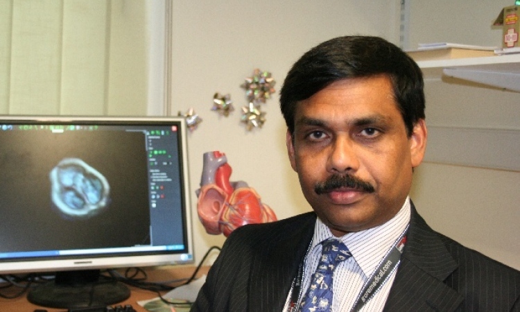
"The aim of the game is opening the chest through little keyholes to operate in the most minimally invasive way possible and avoid sternotomy," said Prof. Friedrich Mohr, Program Director at the Leipzig Heart Center, who review new surgical techniques.
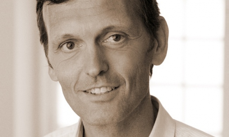
Dr. Mohr was joined by his colleague Dr. Volkmar Falk, Co-Director of the Surgical Department of the Heartcenter who offered a dizzying review of the advances in cardiac surgery with the convergence of medical imaging and powerful computing power.
Citing as an example transcatheter aortic valve implantation (TAVI), which he called the « most exciting tenchological development in the past five years, » Dr. Mohr said this complex procedure demands a team approach between the cardiologists and surgeon, demands a hybrid surgical theater combining the capabilities of a cath lab and open-heart operating room, and also demands advanced imaging, specifically for three-dimensional echocardiography.
Multiple modalities of imaging are required for pre-operattive planning that is essential to determine the precise patient anaomy.
Imaging is again necessary to determine the best route for the transcatheter intervention, whether through the femoral artery, or if these are too restricted, then more directly through the chest wall in a transapical procedure.
« With between 3,000 and 4,000 implantation to date using both types of approaches and different devices, worldwide we have seen results that indicate excellent early outcmes among these patients who are all 75 years or older, » said Dr. Mohr.
Despite the demands and complexity, these new cardiac procedures are improving patient's lives, he said, proudly identifying a member of the audience who wbenefited from a valve replacement procedure.
«Today we perform multiple cases every day in Leipzig, » he said, ading that the Heart Center will record almost 600 patients this year, which is up from 498 patients in 2008 and a 10-fold increase from just 10 years ago at the center.
Dr. Falk attributed the new opportunities for cardiac interventions emerging today to the advances made in neuro surgery where imaging and computing combine for sophisticated planning and intraoperative navigation.
« Today we can not only model preciselythe specific paytient anatomy but the instruments for the intervention as well, » he said.
Moving to the heart, however presents several levels of additional challengesover neuro surgery.
First the heart is a soft tissue suspended in more soft tissue and not as close to hard structures that can be used for navigation.
Second the modeling of the heart requires not only mapping of morphology but rendering images representing perfusion and flow, angiography, and increasing function.
Mapping through imagery the heart function allows surgeons to deterlmine the target and the type of surgery to be performed.
« There are newer imaging modalities that are proving to be indispensible for functional analysis and for surgical guidance, » he said, citing fused SPECT-CT where spectrtal images show perfusion deficits and often reveal clear cut evidence for an intervention.
Dr. Falk also said that fusing CT and MRI data increasing are being used to show perfusion deficits for targeting the intervention.
Other image fusion modalities include combining CT images with electrophysiology data, as well as CT placed on top of the intraoperative angiography.
A novel use of advanced imaging techniques is rapid prototyping for modeling the left ventricle. Using 3-D imaging enables creation of a template for more precise volulme modeling, he said.
The importancee of imaging in cardiac surgery increases the more the surgeon reduces the invasivenes of the procedure, to the point that in endoscopic procedures, « imaging provides the only possible view of the surgery site, » he said.
Two future applications for advanced imaging technique introduce a new partner in the surgical suite, the robot.
A project at the Leipzig Heart Center in partnership with Siemens is investigationg the use of the syngo Dyna CT, a robotically driven fluoroscopy unit.
Originally developed for use in neuro and abdominal surgery, Dr. Falk is using the Dyna CT to generate CT-like data of the heart that can be combined with software capable of detecting diseased tissue.
21.11.2009