Image source: RSNA; from: Xu et al., Radiology 2023
News • Lung cancer, cardiovascular disease
AI uses low-dose CT data for mortality prediction
Artificial intelligence (AI) can use data from low-dose CT scans of the lungs to improve risk prediction for death from lung cancer, cardiovascular disease and other causes, according to a new study.
The research was published in Radiology, a journal of the Radiological Society of North America (RSNA).
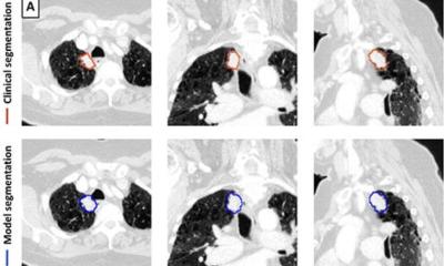
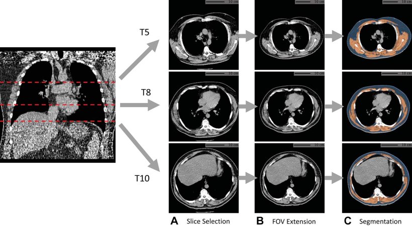
The U.S Preventive Services Task Force recommends annual lung screening with low-dose CT (LDCT) of the chest for individuals ages 50 to 80 years with a high risk of lung cancer, such as longtime smokers. Along with images of the lungs, the scans also provide information about other structures in the chest. “When we’re looking at the CT images, the primary focus is on identifying nodules suspicious for lung cancer, but there is much more anatomical information coded in the space, including information on body composition,” said study lead author Kaiwen Xu, a Ph.D. candidate in the Department of Computer Science at Vanderbilt University in Nashville, Tennessee.
In the space of the chest CT used for lung cancer screening, you can also check other information like body composition or coronary artery calcification that is directly associated with cardiovascular disease risk
Kaiwen Xu
Xu and colleagues previously developed, tested and publicly released an AI algorithm that automatically derives body composition measurements from lung screening LDCT. Body composition is a measure of the percentage of fat, muscle and bone in the body. Abnormal body composition, such as obesity and loss of muscle mass, is linked with chronic health conditions like metabolic disorders. Studies have also shown that body composition is useful in risk stratification and prognosis for cardiovascular disease and chronic obstructive pulmonary disease. In lung cancer therapy, body composition has been shown to affect survival and quality of life.
For the new study, the researcher assessed the added value of the AI-derived body composition measurements. They used the CT scans of more than 20,000 individuals drawn from the National Lung Screening Trial. Results showed that including these measurements improved risk prediction for death from lung cancer, cardiovascular disease and all-cause mortality. “Automatic AI body composition potentially extends the value of lung screening with low-dose CT beyond the early detection of lung cancer,” Xu said. “It can help us identify high-risk individuals for interventions like physical conditioning or lifestyle modifications, even at a very early stage before the onset of disease.”
Measurements associated with fat found within a muscle were particularly strong predictors of mortality—a finding consistent with existing research. Infiltration of skeletal muscle with fat, a condition known as myosteatosis, is now thought to be more predictive for health outcomes than reduced muscle bulk.
The body composition measurements from lung screening LDCT are an example of opportunistic screening when imaging for one purpose provides information about other conditions. The practice is thought to have great potential for routine clinical use. “The images in a CT ordered for quite a different purpose—in our case, early detection of lung cancer—contain much more information,” Xu said. “In the space of the chest CT used for lung cancer screening, you can also check other information like body composition or coronary artery calcification that is directly associated with cardiovascular disease risk.”
The study looked at individuals at a baseline screening only. For future research, the researchers want to perform a study longitudinally; that is, follow the individuals over time to see how changes in the body composition relate to health outcomes.
Source: Radiological Society of North America
26.07.2023