Search for: "brain tissue" - 250 articles found

Photo

Article • ECR 2026 imagines the future of the field

Enhanced by AI, but guided by humans: Radiology’s vision for 2050

Diagnostic imaging without actual images, but with sleek and shiny scanners; no more radiology and pathology departments, but virtual patient models and AI-enhanced surgical precision: At this year's European Congress of Radiology (ECR) in Vienna, two leading experts envisioned 2050 as a radically different future of medicine – less Grey's Anatomy, more Star Trek. They made it clear…

Photo

Article • Developments in brain imaging for psychiatry

Radiologists explore new frontiers of the mind

Psychiatrists and neuroimaging experts gathered to explore one of the last remaining frontiers in radiology – the human mind – at the annual meeting of the French Society of Radiology (JFR) last October in Paris. Their conversation oscillated between neurons and narratives, algorithms and emotions – a vivid reminder that psychiatry is not only about understanding the brain, but about…

Photo

Article • From ablation to neuromodulation

The new era of functional radiosurgery

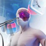
Functional brain radiosurgery is an application of stereotactic radiosurgery (SRS), representing its newest clinical field. It is a precise, non-invasive medical technique using focused ionising radiation to precisely target specific brain structures to modulate brain function for neurological disorders, psychiatric conditions, or intractable pain. The technology offers “precision without…

Photo

Article • On a shoestring budget

Bridging the resource gap for pathology in developing countries

Delivering a pathology service in resource-constrained locations and developing countries remains a challenge. Cost is a significant barrier, as is the availability of equipment, trained staff and technical and IT support can also hinder a desire from clinicians and pathologists to give their patients a high level of service to help their diagnosis and recovery. The subject was tackled in a…

Photo

Article • Precision approach through the eye socket

Orbital gateway to surgical removal of brain tumours

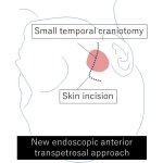
Using a keyhole surgery approach, surgeons have found a new way to access previously difficult-to-reach brain regions with faster recovery times. With the help of 3D modelling technology, the neurosurgeons succeeded in removing complex tumours from the cavernous sinus through the eye socket, avoiding complex brain surgery and enabling their patients to make a quicker recovery.

Photo

News • Hybrid imaging

PET/MR update for added value in theranostics

Bringing diagnostics and therapy closer together - that is the basic concept behind theranostics. Through further development of its PET/MR scanners, Siemens Healthineers aims to advance this approach. Andreas Schneck, Head of the MRI Division at Siemens Healthineers, talks about the new system, which was also presented at the European Congress of Radiology (ECR) in Vienna, and its advantages in…

Photo

Article • Company showcase: Medical Taiwan 2024

Manufacturers from Taiwan present innovative products

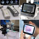
Handheld point-of-care diagnostics, magnetic endoscopy, AI-enhanced robotic surgery, smart patient information management, wireless minimally invasive surgery systems, and much more: At the Medical Taiwan Health & Care Expo in Taipei this summer, visitors had the opportunity to see innovative medical products and solutions across a wide range of specialties. We took a closer look at selected…

Photo

News • Low field imaging

0.05 Tesla MRI: When less is more

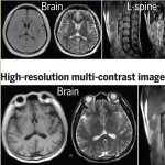
A whole-body MRI scanner with a compact 0.05 Tesla permanent magnet has been developed that operates on a standard wall power outlet without radiofrequency or magnetic shielding cages.

Photo

Article • During lumpectomy

Fluorescence-guided surgery identifies breast cancer in margins

Breast surgery is a traumatic experience for a woman, no matter whether breast-conservation surgery (BCS) or a mastectomy. Trauma levels are greatly enhanced, if pathological evaluation findings of an excised breast tumour following a lumpectomy suggest that additional cancer may still be in the margins, and a second surgical procedure is required. A new system with the ability to accurately…

Photo

Article • Need for diversity in training datasets

Artificial intelligence in healthcare: not always fair

Machine learning and AI are playing an increasingly important role in medicine and healthcare, and not just since ChatGPT. This is especially true in data-intensive specialties such as radiology, pathology or intensive care. The quality of diagnostics and decision-making via AI, however, does not only depend on a sophisticated algorithm but – crucially – on the quality of the training data.

Photo

Article • Flow cytometry

Detecting and measuring nanoplastics in the blood stream

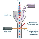
Plastics are a part of everyday life, and an increasingly concerning factor of global environmental pollution. They also have infiltrated our bodies as microparticles (MPs) and nanoparticles (NPs), found even in placentas supporting foetal life. And they are in our blood. Now, researchers in Spain have developed a new method to detect and measure nanoparticles in human peripheral blood that is…
Photo

News • Enabling neuronal tissue growth

A hydrogel to heal the brain

Synthetic hydrogels were shown to provide an effective scaffold for neuronal tissue growth in areas of brain damage, providing a possible approach for brain tissue reconstruction.

Photo

Article • Tissue-engineered glioblastoma model

3D brain tumour in a dish to personalize cancer treatment

It is the size of a common pencil eraser, but it could have a huge impact on the therapy of glioblastoma: Scientists in Virginia have developed a novel 3D tissue-engineered model of the brain tumour microenvironment, which can be used to assess how the glioma cell invades healthy tissue, proliferates, and reacts to chemotherapy drugs.

Photo

Article • Alternative to open surgical procedures gains traction

The future of minimally invasive interventions

In the future, many types of open surgeries will be replaced with minimally invasive interventions, predicts Kevin Cleary, PhD, engineering lead at the Sheikh Zayed Institute for Pediatric Surgical Innovation at Children’s National Hospital, and Professor of Pediatrics and Radiology at George Washington University, both in Washington, D.C. Surgeons and interventional radiologists will be able…

Photo

Article • Benefits of multimodality imaging

Stroke: The importance of workflow

When a patient suffers a stroke, speed in treatment can mean the difference between successful recovery, permanent disability, or death. For Christopher Hess, success in stroke diagnosis is a question of workflow and efficient care delivery.

Photo

Article • Bioprinting

3D biocomposites can repair large bone defects

Loosening hip implants can cause major damage to the bone and a simple replacement won’t suffice to carry the load during movements. “To solve this problem we have to turn to innovative technologies such as bioprinting. Scaffolds are required that – while adapting slowly – offer long-term stability,” says Professor Dr Dieter Wirtz, Director of the Department of Orthopaedics and Trauma…

Photo

Article • Pre-, post- and interoperative

Wearable devices in the surgical environment

Wearable technology has become an important part of medicine, from tracking vital signs to disease diagnosis. In surgery, wearable technologies can now assist, augment, and provide a means of patient assessment before, during and after surgical procedures. Wearable technologies are applied before the patient even reaches the operating room, for example in prehabilitation, i.e. pre-treatment…

Photo

Article • Act fast, save lives

Mobile stroke units: when emergency medicine hits the road

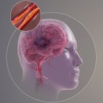
Speed in treatment of ischemic stroke can mean the difference between successful recovery versus permanent disability caused by brain tissue damage or death. Time is of the essence to perform thrombolysis with a tissue plasminogen activate (tPA), a protein that can dissolve blood clots causing the stroke or intra-arterial thrombolytic therapy (IAT) because of large-vessel occlusion.

Photo

News • Coronavirus imaging

Brightest ever X-ray shows lung damage from Covid-19

The damage caused by Covid-19 to the lungs’ smallest blood vessels has been intricately captured using high-energy X-rays emitted by a special type of particle accelerator. Scientists used a new imaging technology called Hierarchical Phase-Contrast Tomography (HiP-CT), to scan donated human organs, including lungs from a Covid-19 donor.

Photo

Article • Neuro- and spine surgery

Perfection in the networked OR: robot, neuro-navigation and VR headsets

At their workplace, neurosurgeons often have to make compromises since most ORs were not designed with the specific needs of their discipline in mind. To address this issue the University Hospital in Essen, Germany, equipped an OR especially for neuro- and spine surgery. The aim is nothing less than revolutionizing the field with the help of digitalisation and cutting-edge technology.

Photo

News • 3D microelectrodes

Promising approach for pain relief without side effects

Researchers at Lund University in Sweden have developed a completely new stimulation method, using ultra-thin microelectrodes, to combat severe pain. This provides effective and personalised pain relief without the common side effects from pain relief drugs. The study, which was conducted on rats, has been published in the research journal Science Advances.

Photo

Article • Mass spectrometry goes handheld

A pen to pin down the fringes of cancer

Mass spectrometry – a powerful tool for analysing the molecular composition of a tissue sample – is invaluable during cancer surgery. However, mass spectrometers are complex and unwieldy, and certainly a poor fit for an operating room (OR). To create a bridge between the lab and OR, Professor Livia S Eberlin, from Baylor College of Medicine, has developed a very special ‘pen’.

Photo

Article • Pregnancy imaging

Machine learning predicts placenta health from MRI scans

Machine learning methods are being used to predict the health of the placenta from a 30-second MRI scan. Researchers hope the approach will offer an insight into the health of expectant mothers and unborn babies by detecting the early signs of dangerous conditions such as pre-eclampsia. Researchers from the School of Biomedical Engineering & Imaging Sciences at King’s College London (KCL)…

Photo

News • Patient response testing

New method predicts which cancer therapies work (and which don't)

A new technology that can study which therapies will work on patients with solid cancerous tumours has been developed by scientists at University College London (UCL). Researchers say the tool, which can rapidly test tumorous tissue against different treatments, such as chemotherapy, immunotherapy or radiotherapy, could be used by clinicians to pinpoint the best therapy for a particular patient.

Photo

Article • Molecular targeting for better results

Emerging novel tracers for cardiovascular imaging

Molecular imaging, guided by novel tracers, is emerging as an important diagnostic and therapeutic tool in cardiovascular medicine. Delegates at ICNC-CT, the online International Conference on Nuclear Cardiology and Cardiac CT, also heard that cardiology can learn from fields such as oncology and neurology that have already made important advances in this area. Professor Frank Bengel, who is…

Photo

News • Brain cancer research

Researchers 3D-print entire active tumor

Researchers at Tel Aviv University have 3D-printed a first-of-its-kind glioblastoma tumor that mimics a living cancer malignancy, powering new methods to improve treatment and accelerate the development of new drugs for the most lethal type of brain cancer. Glioblastoma is notoriously fatal as it accounts for the majority of brain tumors and is highly aggressive. The average survival time of…

Photo

News • Tool to identify tumour mutations

Machine learning fuels personalised cancer medicine

The Biomedical Genomics laboratory at the Institute for Research in Biomedicine (IRB) Barcelona has developed a computational tool that identifies cancer driver mutations for each tumour type. This and other developments produced by the same lab seek to accelerate cancer research and provide tools to help oncologists choose the best treatment for each patient. The study has been published in the…

Photoplay-button-icon

Video • Automated whole-body reperfusion

New technique to increase survival after cardiac arrest

Researchers at the Medical Faculty of the University of Freiburg have developed an improved therapeutic approach to resuscitate people after cardiac arrest - often without neurological complications. Around 50,000 people suffer sudden cardiac arrest in Germany every year. When occurring outside a hospital, the chances of survival are only ten percent. Survivors often suffer from severe permanent…

Photo

Article • Women in medical R&D

Innovation depends on more than just technical skills

Cécile Geneviève is one of the few women who lead research and development (R&D) at a major company and her increasingly female team reflects women’s growing interest in the field. But while gender balance is an important criterion, it takes a broad palette of skills to innovate to alleviate pain for millions of patients, she explained in an interview with Healthcare in Europe.
Photo

News • Preventive potential

Tell-tale signs of heightened stroke risk may appear up to 10 years earlier

The tell-tale signs of a person’s susceptibility to a stroke may appear up to 10 years before the event, suggests research published online in the Journal of Neurology Neurosurgery & Psychiatry. Stroke patients experienced much steeper declines in cognitive abilities and routine daily functioning, starting around a decade before their first stroke, than people who didn’t have a stroke,…

Photo

News • Neuroscience

A deep dive into the brain

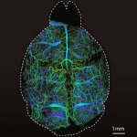
Researchers from ETH Zurich and University of Zurich have developed a new microscopy technique that lights up the brain with high resolution imagery. This allows neuroscientists to study brain functions and ailments more closely and non-​invasively.

Photo

News • Donor organ analysis

New laser technique could improve liver transplant process

Handheld laser devices that help surgeons quickly spot liver damage could transform transplant procedures, research suggests. The non-invasive technique could provide medical staff with instant data on the health of donor livers and help them to identify which organs are suitable for transplant. If widely adopted, the light-based tool could allow more livers to be transplanted safely and…

Photo

News • Understanding myelination

Multiple sclerosis: Mini '3D brains' to speed up research

Tiny 3D models that mimic vital aspects of the human nervous system have been developed in a step that could accelerate drug research for neurological conditions such as multiple sclerosis (MS). The millimetre-wide models – created using stem cells from human skin samples – will be used to study myelin, an insulating substance that helps nerve cells communicate with each other. Researchers…

Photoplay-button-icon

Video • Reparative hydrogel

Fixing traumatic head injury with 'brain glue'

At a cost of $38 billion a year, an estimated 5.3 million people are living with a permanent disability related to traumatic brain injury (TBI) in the United States today, according to the Centers for Disease Control and Prevention. The physical, mental and financial toll of a TBI can be enormous, but new research from the University of Georgia provides promise. In a new study, researchers at…

Subscribe to Newsletter