Image source: Adobe Stock/reineg
News • Genetics & Neuroscience
Improvement of stroke diagnosis and treatment through specific genes
Genes are full of clues about a person's health and might also show the way for stroke recovery. A recent health study at the University of California at Davis (UC Davis) suggests that this may be possible, thanks to time-sensitive gene analysis that allows for faster stroke diagnosis and treatment.
The study has recently been published in BMC Medicine.
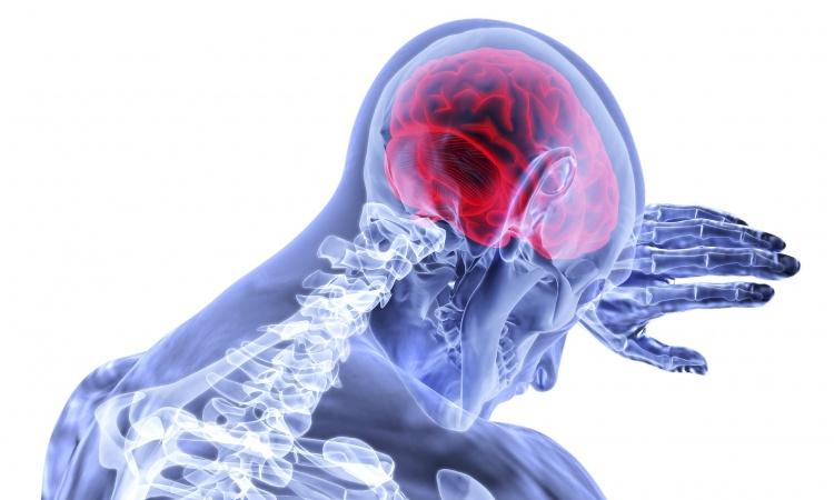
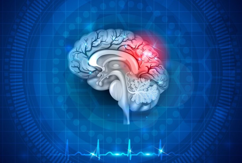
Stroke is the second-leading cause of death and a significant cause of disabilities worldwide. There are two types of strokes: ischemic, caused by a blocked artery, and hemorrhagic, caused by bleeding. During an ischemic stroke, the blood supply to part of the brain is stopped or reduced, preventing brain tissue from getting oxygen and nutrients. The brain responds by swelling, which expands the area of injury. Brain cells begin to die within minutes, leading to increased risk of disability or death.
Successful treatment for strokes currently involves a combination of advanced brain imaging and medication during a short time window. Early diagnosis of a stroke is vital for best outcomes. But some hospitals, especially those which are small or in rural areas or developing countries, may not be equipped with the needed diagnostic technology.
UC Davis Health researchers are trying to determine whether a different treatment option can reliably and accurately result in targeted medication and faster intervention. This treatment involves testing a patient's genes following a stroke.
The results could dramatically change the treatment of strokes
The researchers believe they can analyze the blood of stroke patients to customize treatments to certain genes by isolating specific molecular biomarkers or identifiers that reveal important details about their condition.
"The study results are key to get to the point where we can diagnose stroke without complex imaging, which is not readily available worldwide," said Paulina Carmona-Mora, a research scientist in the Department of Neurology.
"We have identified groups of genes that will allow us to improve diagnosis and identify specific treatments by considering the time after a stroke occurred."
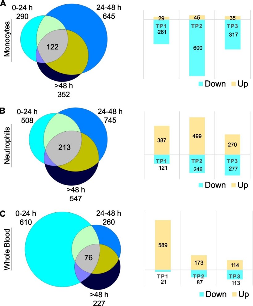
Image source: BMC Medicine (2023). DOI: 10.1186/s12916-023-02766-1
Detailed image description
Differentially expressed genes (DEGs) across time points versus VRFC (0–24 h (TP1), 24–48 h (TP2), and >48 h (TP3)) in monocytes (A), neutrophils (B), and whole blood (C). Venn diagrams of the numbers of DEGs at each time point are shown on the left, and on the right, the corresponding bar plots are shown for the numbers of up- and downregulated DEGs found per time point.
Study focused on cell behavior after stroke
Carmona-Mora and her team profiled the blood of 38 ischemic stroke patients and 18 non-stroke patients who were being treated in hospital emergency departments at the same time. Researchers noted the time elapsed from the onset of the stroke to when the blood sample was collected for the study.
Blood cell samples of the 56 patients were isolated to identify differentially expressed genes, which shows how a cell responds to its changing environment. When the information stored in the cell's DNA is converted into instructions for making proteins or other molecules, it is called gene expression. This acts as an on/off switch to control which proteins are made when they are made and the amounts that are made. Gene expression profiles of stroke patients and the control group of non-stroke patients were analyzed from the blood cell samples.
The study successfully identified gene expression profiles and potential factors that cause changes in gene expression in different blood cell types, which are part of the immune response after stroke.
The researchers detected gene expression that was either abundant, less abundant, or absent at specific times after the stroke. This indicated that many of these genes were associated with stroke severity.
Analysis was performed on the variation of gene expression after stroke at 0–24 hours, 24–48 hours, and more than 48 hours following stroke. This revealed that gene expression in blood cells varies with time after stroke and these changes signal the molecular and cellular responses to brain injury.
Identifying genes and their pathways is critical for understanding how the immune and clotting systems change over time after a stroke. In doing so, this study shows the feasibility of identifying potential time- and cell-specific biomarkers. This would allow researchers to develop targeted therapies most likely to result in a favorable response in each stroke patient.
"Usually, studies use a 'time-agnostic' approach," Carmona-Mora said. "But for this study, examining blood samples in the first couple of days after a stroke was a vital factor. We retained the time information and found which genes can be great biomarker candidates for prompt diagnosis in early stages of the disease. We also found that certain drug targets may be unique to a specific time after stroke, so this can guide treatment in the right time window."
Source: University of California, Davis
19.04.2023


