© Fraunhofer IBMT / Bernd Müller
News • Brain stimulation
Treating neurological diseases with 3D ultrasound
The electrical activity of some 86 billion neurons is what gives the brain the ability to process sensory input, store information, make decisions, and control bodily functions.
This also means diseases and conditions such as Parkinson’s disease, epilepsy, and tremors depend on signal processing and on interaction between the nerve cells. Armed with this knowledge, researchers have spent the past several decades working to treat neurological problems with electric or electromagnetic stimulation of the relevant areas of the brain. But methods such as simulation using external magnetic fields have not yet yielded optimal results owing to the relatively low precision of their effects. Neurosurgery such as in the context of Deep Brain Stimulation (DBS) currently is the clinical standard when it comes to therapeutic brain stimulation. However, it remains a risky procedure due to potential side effects such as hemorraghe or infection.
The ability to precisely target even deep-seated points in the brain and the sequencing of ultrasound signals will open up whole new opportunities for testing and developing individual neurostimulation
Christian Melzer
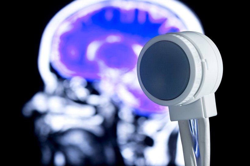
Scientists from Fraunhofer Institute for Biomedical Engineering IBMT in St. Ingbert, in the German state of Saarland, are working on ways to use ultrasound for non-invasive neurostimulation of these areas of the brain. The applicator (an ultrasound probe known as a transducer) is placed on the head using a flexible pad. The ultrasonic signals are of such low intensity that they do not damage the cellular tissue, while at the same time, they can be focused precisely thanks to a function known as 3D beam steering. Medical practitioners and researchers alike have high hopes for the new technology. In the future, it could be used to treat a wide range of neurological diseases and conditions such as epilepsy or to treat the aftereffects of stroke. The Fraunhofer researchers are developing the method as part of various public and industrial research projects, working with partners based in Germany, elsewhere in the EU, and in the U.S., Canada, and Australia.
© Fraunhofer IBMT
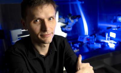
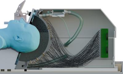
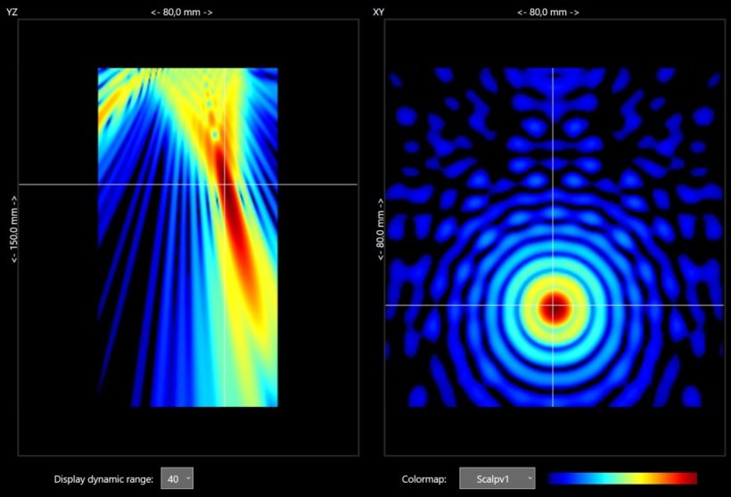
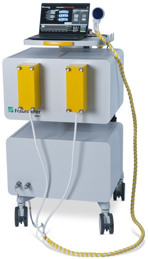
Led by Department Head Steffen Tretbar, the team of Fraunhofer researchers have devised a unique system for the new technology. This approach makes it possible to direct the ultrasonic waves at individual points in the brain and target them even when they are located deep inside the tissue. To achieve this, the team developed a special ultrasound transducer with 256 individual elements, each of which can be controlled individually. Tretbar explains the idea: “Individually controlling the 256 electronic channels makes ultrasound treatment 3D-capable. The transducer elements are arranged like a checkerboard, and they target the desired area of the brain from different angles. That means the focus — the point where the beams meet — can be set to a certain depth in the brain tissue, so the treatment can be tailored to individual patients.”
© Fraunhofer IBMT / Bernd Müller
The Fraunhofer researchers use piezoelectric elements for the transducers. These elements undergo oscillations when voltage is applied, producing ultrasound. The researchers are currently working to further increase precision by using two ultra-sound transducers at once and crossing the beams dynamically in the target area. Combining a very tight focus of between three and five millimeters with practically unlimited placement of the focus deep in the brain makes it possible to precisely modulate the various areas of the brain on a targeted basis without harming the tissue. The ultrasound frequencies used are at the low end of the range, under 1 MHz. For example, a frequency of about 500 kHz may be used. “The person doesn’t feel a thing, and because the ultrasound is low in intensity in the range of what is applied for diagnosis, so that there are no negative side effects on tissue according to the current state of research,” Tretbar explains. There is no need to shave the area for treatment, and medical professionals estimate that a therapy session will take only a few minutes The only preparation needed before the pad with the ultrasound module can be placed on the head is to massage a contact gel into the patient’s hair.
In addition to the ultrasound transducer and electronic components, the team from Fraunhofer IBMT also developed the software used to control the transducer’s 256 individual elements. The software gets the data needed for planning purposes from MRI scans of that patient. The areas of the brain responsible for the patient’s specific neurological problem and their positioning are marked in the image. These markings are then incorporated into a data record that is fed into the control software. The position data can be used to direct the ultrasound signals precisely to where they are needed. It is also possible to program the ultrasound device to transmit the beams in a predefined sequence or follow specific movement patterns. This means doctors could set all of the device’s parameters individually for each patient in the future. “This is still a very new field of research, but it’s also highly promising. Right now, medical centers and researchers all over the world are working to develop and test these kinds of ultrasound sequences,” Tretbar adds.
Fraunhofer IBMT has years of experience in developing ultrasound arrays and multi-channel ultrasound systems and in forming sound beams through beam steering. This expertise has formed the foundation for a universally usable technology platform that is undergoing further development on an ongoing basis. “Researchers can use our technology platform to develop a whole range of different treatments and test them in clinical trials down the road as well,” Tretbar says.
Doctors do not expect ultrasound treatment to cure diseases like Parkinson’s or epilepsy, but they do believe it will have a noticeable effect in alleviating symptoms. Ultrasound is also a promising alternative to conventional medications. In the long run, the new technology could conceivably also be used in scenarios such as breaking up plaque in the brain cells of people with Alzheimer’s disease or to treat depression and addiction disorders caused by neurological factors.
The Fraunhofer team is working with researchers from various project partners and universities. Prof. Andreas Melzer, Executive Director of the Innovation Center Computer Assisted Surgery (ICCAS) at the Leipzig University Faculty of Medicine, has high hopes for the innovative technology: “The ability to precisely target even deep-seated points in the brain and the sequencing of ultrasound signals will open up whole new opportunities for testing and developing individual neurostimulation.”
Source: Fraunhofer Institute for Biomedical Engineering IBMT
17.06.2024


