Image source: University of Copenhagen
News • Quantum sensing prototype
A sensor that detects errors in MRI scans
MRI scanners are used by doctors and healthcare professionals every day to get a unique look into the human body. In particular, they are used to study the brain, vital organs and other soft tissues by way of 3D images of exceptional quality compared to other types of medical imaging.
While this makes the advanced tool invaluable and nearly indispensable for healthcare professionals, there is still room for improvement.
The strong magnetic fields inside MRI scanners have fluctuations that create errors and disturbances in scans. Consequently, these expensive machines (hundreds of Euros per hour) must be calibrated regularly to reduce errors.
There are also special scanning methods, which unfortunately cannot be done in practice today. Among them, so-called spiral sequences that could reduce scanning time, e.g., when diagnosing blood clots, sclerosis and tumors. Spiral sequences would also be an attractive tool in MRI research, where, among other things, they could provide researchers and health professionals with new knowledge about brain diseases. But due to the highly unstable magnetic field, performing these types of scans is not currently an option.
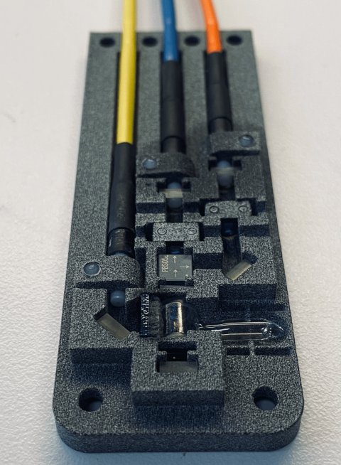
Image source: University of Copenhagen
In theory, the problem can be solved with a sensor that reads and maps changes in the magnetic field. Thereafter, it is relatively simple to correct the errors in images with a computer. In practice, this has been difficult with the current technology, as otherwise suitable sensors interfere with the magnetic field because they are electric and connected to metal cables.
A new invention hopes to make this problem a thing of the past. To combat the problem, a researcher from the Niels Bohr Institute and The Danish Research Centre for Magnetic Resonance (DRCMR) has developed a sensor that uses laser light in fiber cables and a small glass container filled with gas. The prototype is ready and works.
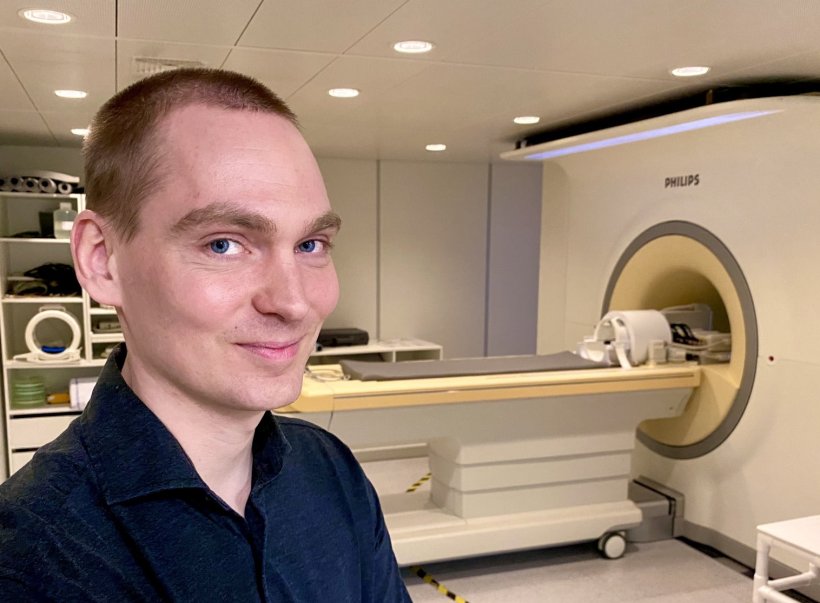
"First we demonstrated that it was theoretically possible, and now we have proven that it can be done in practice. In fact, we now have a prototype that can basically make the measurements needed without disturbing the MRI scanner. It needs to be developed more and fine-tuned, but has the potential to make MRI scans cheaper, better and faster – although not necessarily all three at once," laughs Hans Stærkind, a postdoc at the Niels Bohr Institute and DRCMR at Hvidovre Hospital. Stærkind is the main architect behind the sensor and device that comes with it.
"An MRI scanner can already produce incredible images if one takes their time. But with the help of my sensor, it is imaginable to use the same amount of time to produce even better imagery – or spend less time and still get the same quality as today. A third scenario could be to build a cheaper scanner that, despite a few errors, could still deliver decent image quality with the help of my sensor," says the researcher.
MRI scanners use powerful magnets to produce a strong magnetic field that forces protons in the body's water, carbohydrates and proteins to align themselves with the magnetic field. When radio waves are pulsed through a patient, the protons are stimulated and temporarily spin out of that equilibrium. When they subsequently return to alignment with the magnetic field, they release radio waves that can be used to form real-time 3D images of whatever is being scanned.
Recommended article

Article • Focus on radiology
Magnetic resonance imaging (MRI)
Imaging without ionising radiation: MRI uses magnetic fields to look inside the body. Keep up-to-date with the latest research news, medical applications, and background information on MR imaging.
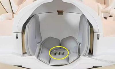
Hans Stærkind's prototype works using a device for sending and receiving laser light that looks like a 1990’s stereo system. It sends laser light through fiber optic cables – i.e., without any metal – and into four sensors located in the scanner.
We can measure the strength of the magnetic field by finding out what the right frequency is. This happens completely automatically and lightning fast by the receiving device
Hans Stærkind
Within the sensors, the light passes through a small glass container containing a caesium gas, which absorbs the light at the right light frequencies. "When the laser has just the right frequency while passing through the gas, there is a resonance between the waves of light and electrons in the caesium atoms. But the frequency – or wavelength – at which this happens changes when the gas is exposed to a magnetic field. In this way, we can measure the strength of the magnetic field by finding out what the right frequency is. This happens completely automatically and lightning fast by the receiving device," explains the researcher.
As disturbances in an MRI scanner's ultra-powerful magnetic field occur, Hans Stærkind's prototype maps where in the magnetic field they are occurring and by what strength the field has changed. In the near future, this could mean that disturbed and faulty images could be corrected – based on the data collected by the sensors, and subsequently made accurate and entirely usable.
The prototype is currently housed at DRCMR at Hvidovre Hospital in Copenhagen, which is also where the idea was conceived. "The original idea came from my supervisor here at DRCMR, Esben Petersen, who is unfortunately no longer with us. He saw huge potential in developing a sensor based on lasers and gas that would be able to measure the magnetic fields without disturbing them," says Hans Stærkind.
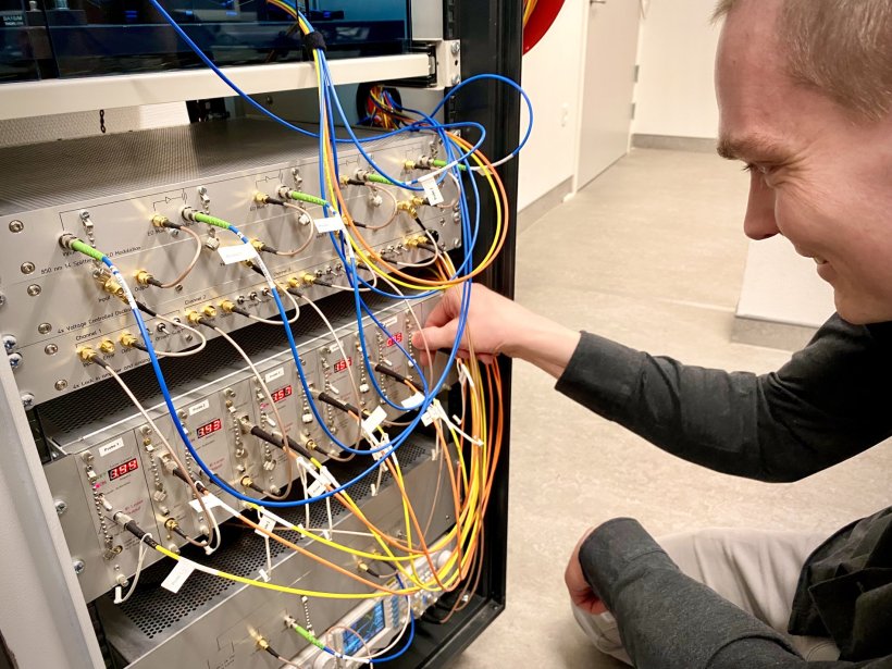
Image source: University of Copenhagen
With the help of quantum physicists at the Niels Bohr Institute, including Professor Eugene Polzik, Stærkind developed the idea into an actual theory. And with the prototype, he has now put that theory into practice. "The prototype is designed in such a way that it is already suitable in hospital contexts as a robust and well-functioning instrument. And so far, our tests have shown that it works as it should. One can imagine that this invention will eventually be integrated directly into new MRI scanners," says Stærkind.
For now, the prototype will be developed further so that its measurements become even more accurate. "We need to collect data and fine-tune it so that it continuously becomes a better and better tool for finding errors in scans. After that, we’ll move on to the exciting work of correcting errors in MRI images, and find out in what situations and which types of scans our sensor can make a significant difference," says the researcher.
According to Stærkind, the immediate target group for his sensor are MRI research units. But he also hopes that one of the large MRI manufacturers finds out about the new technology, in the slightly longer term. "Once the prototype has been refined in a 2.0 version and its qualities documented with plenty of data from actual scans here at the hospital, we will see where this goes. It certainly has the potential to improve MRI scans in a unique way that can benefit doctors and, not least, patients," says the researcher.
Source: University of Copenhagen
04.05.2024


