Image source: University of Waterloo
News • Correlated diffusion imaging
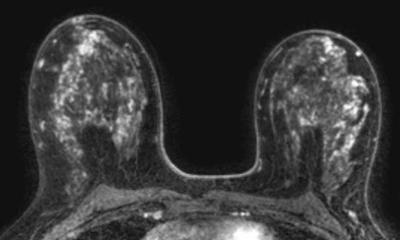
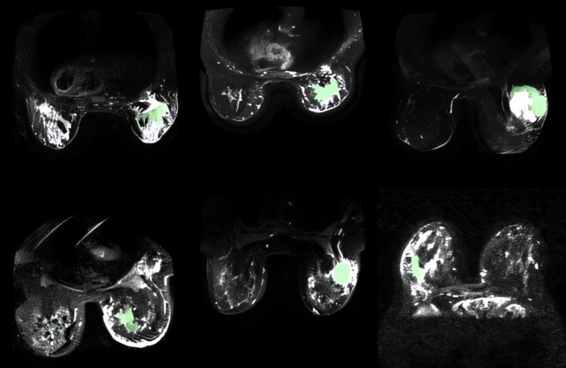
AI makes breast cancer “glow” in MRI images
Imaging technology developed and refined at the University of Waterloo promises better detection and treatment of breast cancer by more accurately pinpointing cancerous tissue.
First successfully applied to prostate cancer, the new form of magnetic resonance imaging (MRI) has now been optimized using artificial intelligence (AI) to make cancerous tissue appear to light up or glow next to healthy breast tissue in images. “This technology has great potential to not only improve breast cancer detection, but also treatment,” said Dr. Alexander Wong, a professor in the Department of Systems Design Engineering at the University of Waterloo and co-director of the Vision and Image Processing (VIP) Lab. “Our images contain key predictive information to help clinicians determine the best courses of action for treating each patient.”
The researchers published their findings in the journal Sensors.
The more accurate we make CDI for delineating between cancerous tissue and healthy tissue, the more effective patient treatment plans and treatment itself can be
Amy Tai
The system works by leveraging specific physical characteristics of breast tissue such as density, and how the irregular packing of cells leads to differences in the way water molecules move in cancerous tissue compared to healthy tissue. Known as synthetic correlated diffusion imaging (CDI), the new technology highlights those differences by capturing, synthesizing and mixing MRI signals at different gradient pulse strengths and timings. The result is significantly better delineation of cancerous breast tissue, making it a potentially powerful tool for doctors and radiologists. “The more accurate we make CDI for delineating between cancerous tissue and healthy tissue, the more effective patient treatment plans and treatment itself can be,” said Amy Tai, a Waterloo engineering PhD student who is supervised by Wong at the VIP Lab.
By giving surgeons more precise information on the margins of tumours, CDI images could help them carefully limit the amount of tissue removed, or ensure all cancerous tissue is removed the first time so a second operation isn’t required. Tailoring the underlying system for breast cancer involved the use of pre-treatment images of more than 350 patients at 10 medical institutions in a study by the American College of Radiology Imaging Network.
Researchers now hope to expand the use of their imaging technology to other kinds of cancer, especially cancers of the neck and head, such as brain cancer. “We have already illustrated great potential for prostate cancer and now we are seeing promising results for breast cancer,” said Wong. “It’s extremely encouraging that we’ll be able to expand and help in other areas as well.”
Source: University of Waterloo
25.02.2025