Image credit: Katsumasa Fujita, Osaka University
News • High-throughput chemical imaging
Acceleration brings Raman spectroscopy within reach of clinical application
Researchers have developed a Raman microscope that can acquire information hundreds of times faster than a conventional Raman microscope. Raman microscopy is a powerful non-invasive tool for performing complex chemical analysis of cells and tissues, and this technology development could help expand its usefulness in biomedical applications.
“Our high-throughput Raman spectral imaging can quickly image and analyze a large area without any sample pretreatment, which could make it useful for medical diagnoses and the tests used to screen for new drugs,” said research team leader Katsumasa Fujita from Osaka University. “The label-free, high-throughput multiplex chemical imaging and analysis enabled by the technique could also be used to enable new applications or overcome limitations of current methods.”
Label-free molecular analysis with Raman imaging would also be useful for efficiently detecting drug response of cells, aiding in drug development
Katsumasa Fujita
In the Optica Publishing Group journal Biomedical Optics Express, the researchers describe their new multiline illumination confocal Raman microscopy approach. It works by detecting separate regions of the sample in parallel, enabling fast Raman hyperspectral imaging. They show that the technique can acquire hyperspectral images of biological tissue with a field of view of 1380 x 800 pixels in about 11 minutes. This would require days to acquire with a traditional Raman microscope. “We hope that high-throughput Raman imaging will eventually make it possible to perform medical diagnoses more efficiently and accurately while possibly enabling diagnoses that weren’t possible before,” said Fujita. “Label-free molecular analysis with Raman imaging would also be useful for efficiently detecting drug response of cells, aiding in drug development.”
Raman spectroscopy provides important insights into the chemical makeup of a sample by using light to excite molecular vibration. The resulting molecular vibrations create a type of chemical fingerprint that can be used to identify the sample’s composition. Raman microscopy takes this one step further by obtaining very high-resolution spectral images, which are useful for imaging cells and tissues. However, due to the tradeoff between spectral resolution and imaging speed, Raman microscopy hasn’t been practical for use in the clinic.
The new multiline illumination approach builds upon a technique the research team previously developed known as line-illumination Raman microscopy. That approach was faster than conventional confocal Raman microscopy and enabled dynamic imaging of living cells but was still too slow for the large-area imaging often required for medical diagnosis and tissue analysis. “To address this issue, we developed multiline illumination Raman microscopy, which acquires large-area images about 20 times faster than line-illumination Raman microscopy,” said Fujita. “With our new technique, the spectral pixel number – or resolution – and imaging speed can be adjusted, depending on the application. In the future, even faster imaging speed might be possible as cameras continue to be developed with more pixels.”
Recommended article
Article • Surface-enhanced Raman spectroscopy
The lab-on-a-chip SERS platform
Analytically sensitive and specific detection of pharmaceuticals or metabolites in bodily fluids, as well as fast and reliable detection of human pathogens, are major challenges for instrument-based analytics in medical diagnostics. Over the past few years the combination of surface-enhanced Raman spectroscopy (SERS) and microfluidic devices (Lab-on-a-Chip) has emerged as a perfectly suited…
The team’s new multiline-illumination Raman microscope irradiates about 20,000 points in a sample simultaneously with multiple line-shaped laser beams. The Raman scattering spectra generated from the irradiated positions are then recorded in a single exposure that contains the spatial information for the Raman spectra in the sample. Scanning the laser beams across the sample allows a two-dimensional hyperspectral Raman image to be reconstructed. To accomplish this, the researchers use a cylindrical lens array — an optical element composed of periodically aligned multiple cylindrical lenses — to generate multiple line-shaped laser beams from a single laser beam. They combined this with a spectrophotometer capable of acquiring 20,000 spectra at the same time. Optical filters were also important for avoiding cross talk among the spectra at the spectrophotometer detector.
A high-sensitivity, low-noise CCD camera with a large number of pixels was also critical. “This CCD camera allowed 20,000 Raman spectra to be distributed on the CCD chip and detected simultaneously,” said Fujita. “The custom-made spectrophotometer also played an important role by forming the 2D distribution of spectra on the camera without significant distortion.”
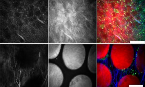
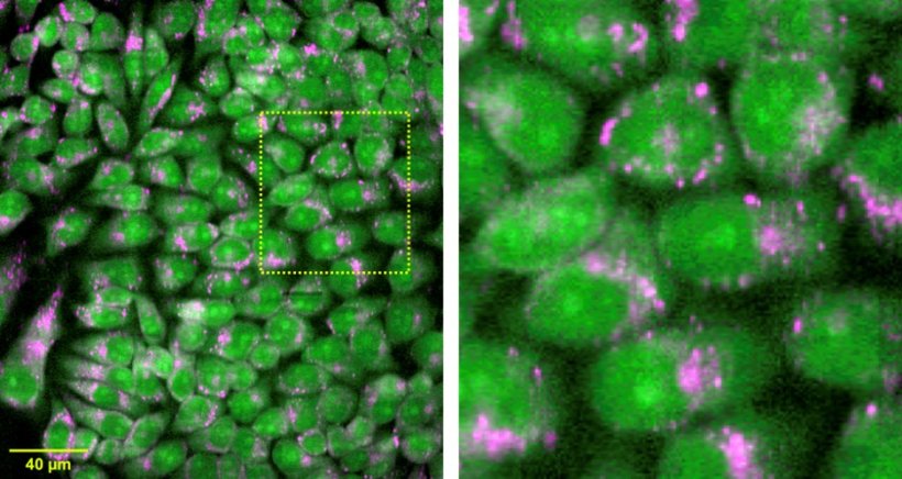
The researchers used the new technique to acquire measurements from live cells and tissues to test its imaging performance and potential in biomedical applications. They showed that irradiating a mouse brain sample with 21 simultaneous illumination lines could be used to acquire 1,108,800 spectra in just 11.4 minutes. They also performed measurements on mouse kidney and liver tissue and conducted label-free live-cell molecular imaging. “Small-molecule imaging and super-multiplex imaging using Raman tags and probes could also benefit from this technique because they don’t require a large number of pixels in a spectrum and can benefit from fast imaging,” said Fujita.
For this technique to be applied for medical diagnoses, the researchers say it would be important to build a database of Raman images, something that can be accomplished efficiently with the new Raman microscope thanks to its speed and large imaging area. They are also working to increase the system’s speed by a factor of about 10 and would like to reduce the cost of camera, laser, and spectrophotometer to make commercialization more practical.
Source: Optica Publishing Group
09.02.2023